There is a change in the clinical manifestation of Parkinson’s disease (PD) over time. Figure 1 provides a schematic overview of this process, starting with a pre-symptomatic phase, which lasts for approximately seven years. Following that, for anything from six to 15 years, patients develop symptoms and complications, but these respond very well to dopaminergic drugs and surgical intervention. This early stage of dopaminergic deficit has been the subject of the most attention over the past decades and, as a result, a range of therapeutic options now exist.
In late-stage PD, which more patients reach as their life expectancy increases, other problems begin to manifest, including cognitive decline, dementia and equilibrium problems, which are theorised to relate to a wider pathology. This stage can last for 10–15 years or more, and there are currently few treatment options available. Understanding the underlying structural changes in the brain that drive this evolution is essential.
Pathology of Parkinson’s Disease
The basal ganglia, in simple terms, comprises two systems. The first is the nigrostriatal dopaminergic projection, which is a dopaminedependent modulator of corticostriatal and thalamostriatal afferent. The second is the subthalamic glutamatergic circuitry, which modulates the globus pallidus pars externa (GPe) and globus pallidus pars interna (GPi) and therefore the output of the basal ganglia, which feeds back to the thalamus and cortex/brainstem. These two systems effectively control much of the motor state of a patient. Denervation in this section of the brain reduces dopamine modulation and hence motor control. This results in the typical PD fluctuations from ‘off’ to ‘on’ states with characteristic patterns, including biphasic dyskinesias at the beginning and end of a dose and levodopa-induced dyskinesias during the on state.1
The remarkable effect of deep-brain stimulation of the subthalamic nucleus (STN-DBS) on motor fluctuations in parkinsonian patients, even in the absence of other medication, emphasises the importance of the nigrostriatal and globus pallidus systems of the basal ganglia in terms of motor control. Despite the intra-patient variation, the overall success of STN-DBS demonstrates the power of modulating these systems. Even a very small stimulation in a highly localised area of the basal ganglia can turn tremor into dyskinesia. Therefore, the motor system in PD can be explained simply by modulation of these two systems in the basal ganglia: dopamine in the striatum, particularly in the putamen, and activity in the STN modulating basal ganglia output. This is a remarkable arrangement and it is unlikely that there is another target as good as the basal ganglia and its subsystems for modifying motor activity. The Antiparkinsonian Armamentarium
Many dopaminergic drugs and treatments are currently available in several different classes: orally available medications, namely levodopa and dopamine agonists; continuous infusions of levodopa/carbidopa or apomorphine; and bilateral surgery of the STN or GPi. Subcutaneous apomorphine has been used for more than 20 years, and was first tested in the UK by Andrew Lees and his team.2 This is approximately contemporary with the first recorded example of the effect of continuous levodopa infusion, by Jorge Juncos et al. in 1985.3 However, unlike with apomorphine, it took a long time to make continuous levodopa infusions a practical reality.
Consequently, with all of these therapeutic options available, it is fair to say that replacing the dopamine deficit is no longer a major challenge in PD. The main problem now is to try to control what happens to patients after the first 10–20 years of their disease in the advanced phases (see Table 1). This is where problems such as sleepiness, falling, autonomic disturbance and cognitive decline start to manifest, and this is where the major challenge lies for the future.
In order to study the progression of PD, it is essential to understand the changes in the brain and what effect these have on patients and their quality of life. After some 12–15 years of evolution, mild cognitive impairment (MCI) and dementia become more prevalent in PD. Patients start to exhibit symptoms of executive dysfunction such as abnormal planification and difficulties in performing activities that require temporal ordering (which tend to be subclinical). As the condition progresses, patients also start exhibiting symptoms of attention deficit, lack of verbal fluidity, visuo-spatial dysfunction and further executive dysfunction, such as somnolence, disorientation and problems with semantic memory.
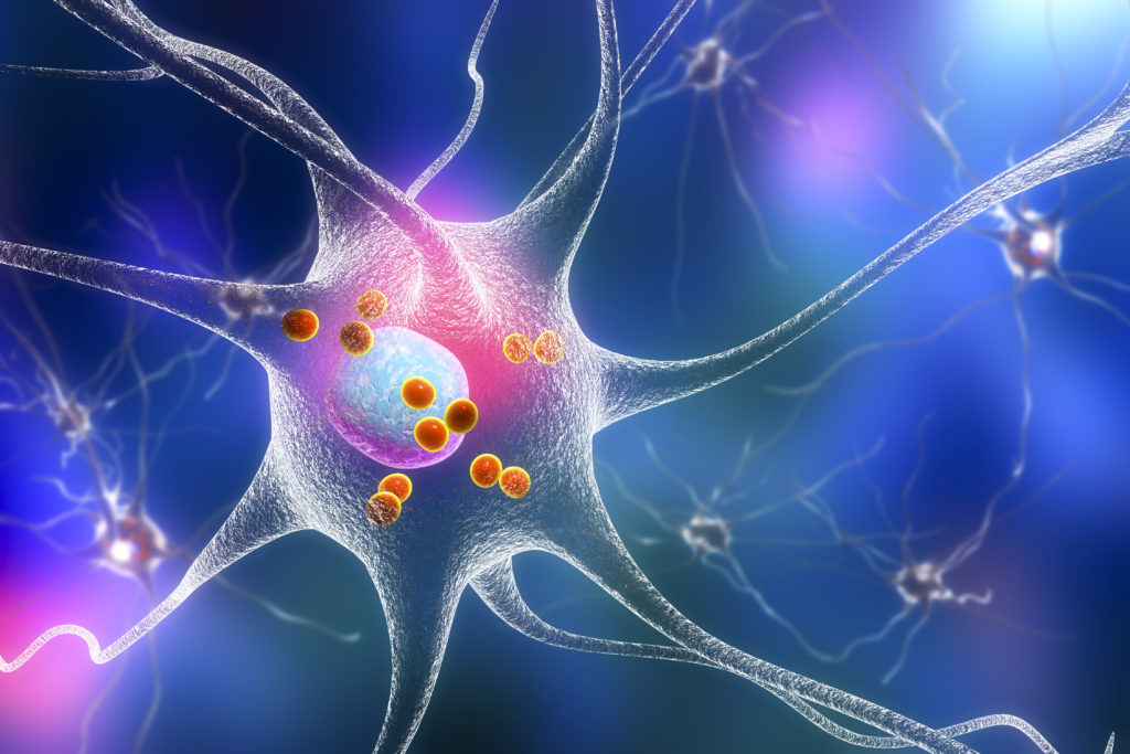
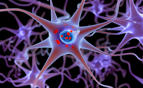
In terms of the pathology, as PD advances, characteristic aggregates of alpha-synuclein – Lewy bodies – start to spread. In the amygdala and the ventral temporal cortex, the presence of Lewy bodies is correlated with visual hallucinations.4 Thus, as PD progresses and the alphasynuclein deposits spread beyond the substantia nigra to affect other brain areas, numerous new problems emerge. Such late-stage effects were not apparent before the 1980s, when the major issues connected with PD were severe off states and dystonia, etc. There is currently no way to halt or reverse the progression of PD. New strategies have been developed to help patients and families and provide support and rehabilitation, but these palliative approaches recognise that as yet there is no long-term solution available.
Magnetic Resonance Imaging Evidence
To try to ascertain the extent and severity of this evolution in PD, we undertook a study with a number of patients with cognitiveimpairment using magnetic resonance imaging (MRI). It is known that there are differences between the MRI scans of patients with earlystage PD, those with MCI and those with dementia, but this study attempted to go further and explore the size and extent of these changes. In this study we found that demented patients showed wider and more intense atrophy than either patients with MCI or the control group of PD patients with no cognitive impairment. The differences between the scans of the control group and the group with MCI were minor in comparison.5 This implies that MCI is an earlier state than dementia from which some patients will progress but others may not.
A similar study undertaken in the US used positron emission tomography (PET) to investigate the differences in metabolism and glucose uptake in the PD brain, and thereby determine activity in the striatum and thalamus. The US researchers found that the brains of PD patients with dementia show evidence of hypometablism in the occipital cortex and some frontal regions. This contrasts with the hypermetabolism seen in the basal ganglia of PD patients, particularly in the motor loop of the posterior putamen. Analysing these images,the researchers found statistical differences in the pattern of abnormalities observed in the scans of demented brains. Once again, a comparison of matched controls (with PD but without MCI) and patients with MCI showed the same evidence of hypometabolism, but in much smaller and more focused areas. The researchers speculated that quantification of these alterations using PET may allow for objective assessment of the progression and treatment of PD.6
Therefore, there is hope that this early phase of MCI in PD is not necessarily associated with profound cell loss or cell dysfunction. There may be an ‘oil drop’ pattern of evolution of PD, where the focal point spreads out over time to other regions. By intervening surgically or therapeutically at one particular focal point it may be possible to halt progression before full dementia is reached.
Future Research Avenues
There are many groups still working to further improve dopaminergic therapies. While this line of research is welcome, it is no longer essential because there are now many treatment options for the early and intermediate phases of PD. Another avenue of exploration is to identify new PD drugs that work on non-dopaminergic targets. For example, such drugs might affect adenosine modulation in the basal ganglia or acetylcholine activity in the cortex. The results of such a strategy are currently unpredictable, but may be worth pursuing. Other approaches that also have the aim of replacing striatal dopamine deficiency, such as cell therapy, are unlikely to have a great future either. Far more important is the development of agents that can modify the evolution of PD. If PD could be stopped at the time of diagnosis, its evolution could become much more benign. Gene therapy potentially falls into this category, as it is dedicated to changing the neurodegeneration process, modifying disease and making a long-term difference, although any clinical application of this technology is still many years away.
The future expectation is to find, at the molecular level, a common celldeath mechanism that may be blocked or restored; a cellular equivalent of the dopamine system or the glutamatergic system in the basal ganglia. This includes understanding the role of Lewy bodies – whether they are a protective reaction or whether they are deleterious, or both.
Three publications in Nature Medicine in early 2008 added more interesting data to the debate on the pathology of PD. All three concerned PD patients who, between nine and 16 years ago, had received foetal cell grafts in their striatum. This means that while the patients were each around 70 years old, the cells in their grafts were substantially younger. Through post mortem examinations, two of the three groups of researchers found Lewy body inclusions in the foetal cell grafts.7,8
Although the third group did not report the same finding,9 it can be said that an absence of an abnormality is less compelling evidence than its presence; not all brains would be expected to display identical pathology. The two positive Lewy body results lend weight to the idea that PD is a process that is always present – either in the environment or as a pathological process in the brain – rather than the result of a single insult to the brain that slowly evolves, for example, as with prion disease. Under this hypothesis, the striatum cannot be considered the primary site of pathology but rather the primary region to be attacked by the degenerative process, where Lewy bodies represent the expression of the underlying pathology.
Summary and Conclusions
The last 20 years have seen a great move forwards in terms of available treatments for the early and intermediate stages of PD. As patients have benefited from this enlargement of the armamentarium and improvement in management strategies, thereby extending their lifespan, new problems have presented themselves. Most notably, these include the decline to dementia, which further erodes a patient’s quality of life. The next step, therefore, is to better understand the underlying disease mechanism in Parkinson’s disease to try to work out a way to halt or at least slow it down. It is likely that the two systems of the basal ganglia provide the best opportunity for intervention: while advanced Parkinson’s disease exhibits a wider disease pattern, the focal point remains in this region. The role of Lewy bodies is still largely uncertain; the latest data provide a clue to the nature of the processes underlying Parkinson’s disease, but also serve as a warning that this is a complex issue. The dream of finding a common celldeath mechanism is still many years away from being fulfilled.

