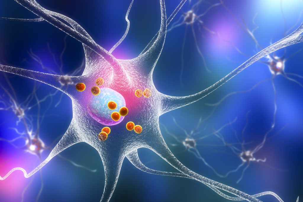
Parkinson’s disease (PD) is caused by a progressive degeneration of dopaminergic cells in the pars compacta of the substantia nigra (SNc), thereby inducing a depletion of dopamine concentration at striatal level. The restoration of dopaminergic transmission by levodopa (L-dopa) has been used successfully for many years, but causes long-term motor complications. In this case, neurosurgery has the potential to help restore the motor function of patients.
Experimental Origin of Subthalamic Stimulation
During the last decade, the subthalamic nucleus (STN) has become a target of choice for the surgical treatment of severe forms of PD. Its implication in the pathophysiology of the disease was originally determined from experimental data in animal models. Numerous experimental studies have demonstrated that the depletion of dopamine induces a disorganisation of neuronal activity in basal ganglia structures. In rodent (6-hydroxy-dopamine (6-OHDA)-lesioned rat) and non-human primate (monkeys rendered parkinsonian by systemic injections of 1-methyl-4-phenyl-1,2,3,6-tetra-hydro- pyridine (MPTP)) models of PD, abnormal activity of STN neurons has been reported. Increased firing rates and/or important changes in the firing pattern of STN neurons have also been shown. According to the generally accepted model of the functional organisation of basal ganglia,1 these changes result in hyperactivity of the output structures of basal ganglia, the pars reticulata of the substantia nigra (SNr) and the internal part of globus pallidus (GPi). In this way, an increase in the tonic inhibitory influence exerted by these structures on the motor thalamic nuclei results in a deactivation of motor cortical areas.
At the beginning of the 1990s, the STN was identified as a potential target for neurosurgical therapy for PD. STN lesions in MPTP-treated monkeys induces an improvement in parkinsonian motor symptoms that accompanies the expression of abnormal movements.2 Due to the fact that STN lesioning is irreversible, the author and his colleagues have proposed to replace this approach by electrical high-frequency stimulation (HFS). This research group has shown that HFS (100Hz frequency) of the STN alleviates parkinsonian motor symptoms in monkeys rendered parkinsonian by MPTP, without any evident side effects. Quantification of motor performance has shown that HFS of the STN induces a normalisation of both reaction and movement times as well as electro-myogram (EMG) activity of agonist/antagonist muscles.3 This beneficial effect is comparable with that obtained after the administration of L-dopa without motor fluctuations. Based on such experimental evidence, HFS of the STN has become the most promising therapy for the treatment of PD.
Clinical Effect of Subthalamic Stimulation
Since 1993, HFS of the STN has been successfully transferred to human patients and has been shown to alleviate all the major motor symptoms of PD while allowing a dramatic reduction in daily levodopa (L-dopa) requirements and dyskinesias.4 Bilateral STN HFS (130Hz, 0.06 millisecond (ms) pulse width at 2 to 4 volts) improves parkinsonism considerably more than unilateral STN deep brain stimulation (DBS).5 Krack et al.6 suggested that parkinsonian patients who do not respond well to L-dopa treatment or in whom a post-synaptic dopaminergic lesion are not good candidates for STN surgery. The Unified Parkinson’s Disease Rating Scale (UPDRS) motor examination scoring after STN HFS is decreased by approximately 60%. Motor fluctuations also tend to disappear and off-period dystonia is immediately alleviated. Moreover, treated patients no longer need help in their daily activities. Bilateral STN stimulation improves most axial features that responded to L-dopa before surgery.7
Recent work has shown that the efficacy of STN HFS in reducing motor symptoms and L-dopa-induced dyskinesia in patients with severe PD is largely maintained for five years after surgery.8 Daily living activity (UPDRS II) was dramatically improved by STN HFS. Parkinsonian motor disability (UPDRS III) was also improved. L-dopa daily doses were reduced by 58%. The score for speech only improved during the first year and then progressively worsened, returning to baseline levels at five years after surgery. However, with time, there is deterioration in akinesia, axial symptoms, and cognitive problems associated with the progression of the underlying disease. Stimulation of the STN also seems most useful for relatively young patients who have motor complications from L-dopa treatment and who are independent in terms of daily living activities in their best on-medication state. Examination of the long-term effects of STN HFS has shown that despite motor and cognitive decline, the marked improvement in motor performance and daily quality of life was maintained five years after surgery.
Mechanisms of HFS of the STN
Despite the growing clinical experience with STN HFS as a therapeutical neurosurgery approach of PD, its mechanisms of action are not yet fully understood. The observation that HFS of the STN mostly mimics the STN lesion effects has suggested that this type of stimulation acts by silencing STN neurons and subsequently reduces their excitatory influence on the output structures of the basal ganglia. In keeping with this hypothesis, in vivo electrophysiological recordings from normal and 6-OHDA-lesioned rats have shown that STN HFS decreases the activity of STN neurons.9,10 Consistent with this effect, metabolic data have shown a significant decrease in the levels of cytochrome oxidase subunit I (CoI) messenger (m)RNA expression in the STN in both normal and 6-OHDA-lesioned rats.10–12 In rat brain slices, it has been shown that HFS induces a suppression of subthalamic activity13 or has a dual effect, i.e. spontaneous activity is silenced and replaced by a new type of burst activity, suggesting a direct activation of voltage-sensitive membrane currents in the cell bodies and axons of STN neurons.14 In human patients during functional stereotaxic surgery, HFS induces inhibition of STN neuronal firing.15,16 A more recent study in MPTP-treated monkeys from the author’s group has provided new evidence that STN HFS decreases firing rate and triggers abnormal oscillatory activity in the STN nucleus.17 As abnormal synchronised oscillations are supposed to be, at least partially, at the origin of the parkinsonian syndrome, the reduction of oscillations might be as important as the impact on firing rate for the beneficial effects of STN stimulation.
Although HFS is believed to inhibit the activity of STN neurons, there is much conflicting data as to whether this type of stimulation inhibits or excites STN efferent structures. In 6-OHDA rats, the author’s group reported that STN HFS produces an inhibitory effect on the majority of SNr and entopeduncular (the rodent equivalent of GPi) neurons.9,10,18 Maurice et al.19 observed a decrease in SNr activity when STN HFS was performed with low intensity and an increase in activity when the STN was stimulated at higher intensity, suggesting that a combination of distinct effects on somatic and axonal activity might occur above a certain intensity threshold. It has been suggested that the inhibitory effect of STN HFS is due to the excitation of gamma-aminobutyric acid (GABA)- ergic fibers originating from the globus pallidus. A microdialysis study carried out in 6-OHDA rats has shown that STN HFS induces an increase in GABA levels in the SNr without any change in extracellular glutamate level.20 In contrast, Hashimoto et al.21 observed that in MPTP-treated monkeys, STN HFS increases the mean firing rate of GPi neurons and modifies their firing patterns from spontaneously irregular to regular discharge, suggesting that stimulation might excite STN neurons or STN efferents that project to GPi. These paradoxical experimental results might be explained by the model proposed by McIntyre et al.22 showing that sub-threshold stimulation results in a suppression of intrinsic firing activity through activation of pre-synaptic terminals and that suprathreshold stimulation suppresses somatic intrinsic firing while generating efferent output via activation.
Conclusion
In conclusion, it is now accepted that HFS of the STN represents an effective clinical therapy for severe forms of PD, even though the mechanisms of action are not fully understood. On account of the adaptability and reversibility of the technique, HFS of other brain structures is successfully used in other pathologies, such as the GPi for dystonia and the nucleus accumbens for obsessive convulsive dis-orders, among others. ■

