touchNEUROLOGY touchNEUROLOGY
Movement Disorders, Parkinson's Disease
Read Time: 9 mins

New Magnetic Resonance Imaging Biomarkers Advance the Characterization of Parkinson Disease

Copy Link
Published Online: Nov 2nd 2013 US Neurology, 2013;9(1):8–12 DOI: http://doi.org/10.17925/USN.2013.09.01.8
Authors: David A Ziegler, Suzanne Corkin
Quick Links:
Abstract
Article
Article Information
Abstract:
Overview

The pathophysiology of idiopathic Parkinson disease (PD) is traditionally characterized as substantia nigra degeneration, but careful examination of the widespread neuropathologic changes suggests individual differences in neuronal vulnerability. A major limitation to studies of disease progression in PD has been that conventional MRI techniques provide relatively poor contrast for the structures that are affected by the disease, and thus are not typically used in experimental or clinical studies. Here, we review the current state of structural MRI as applied to the analysis of the PD brain. We also describe a new multispectral MRI method that provides improved contrast for the substantia nigra and basal forebrain, which we recently used to show that these structures display different trajectories of volume loss early in the disease.

Keywords

Parkinson’s disease, MRI, substantia nigra, basal forebrain, neurodegeneration, biomarkers

Article:

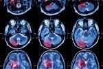
Parkinson disease (PD) is a devastating neurodegenerative disorder Characterized by its cardinal motor symptoms: resting tremor, muscular rigidity, bradykinesia, postural instability, and gait abnormality.1 PD currently affects 1–2 % of individuals over age 65, totaling five million people worldwide. During the next 20 years, the incidence of PD is projected to double, making research on its causes and treatments timelier and more relevant to global public health than ever before. A major impediment to such research is a paucity of safe, fast, and effective brain imaging methods for visualizing the structures affected by PD. Because conventional structural magnetic resonance imaging (MRI) techniques cannot visualize the brain changes that are at the core of this disease, MRI-based biomarkers for diagnosis and tracking disease progression do not currently exist.

The cardinal motor features of PD are typically attributed to a loss of nigrostriatal dopaminergic neurons in the substantia nigra pars compacta (SNpc), which is accompanied by the aggregation of Lewy bodies and neurites in this structure.2–4 While denervation of dopaminergic nigrostriatal projections may explain the primary motor symptoms of PD, as shown by the dramatic motor improvement associated with dopamine replacement therapy,5,6 abnormalities beyond the SNpc7–10 likely underlie the serious and potentially debilitating non-motor features, including cognitive and memory impairments and progression to dementia.11,12 Notably, degeneration of the cholinergic basal forebrain (BF)13–15 and noradrenergic locus coeruleus16,17 in PD probably contribute to non-motor deficits. Although deterioration in the BF and LC is most often associated with late-stage PD with dementia,18 subtle changes in earlier stages could result in poor performance on tests of memory and attention.19 Research on these non-motor aspects of the disease has been hindered by a lack of sensitive MRI biomarkers for the affected structures. This article reviews recent progress in developing new MRI-based biomarkers to visualize and characterize abnormalities in some of the brain structures affected by PD.

Imaging the Substantia Nigra
The substantia nigra is comprised of two structurally and functionally segregated regions: the SNpc, which projects mainly to the striatum and basal ganglia, and the substantia nigra pars reticulata (SNr), which sends its primary efferent projections to the thalamus and superior colliculus. In PD, neuronal loss in the SNpc is prevalent in the caudal and mediolateral part and more limited in mediorostral areas.20 This loss of SNpc neurons results in a marked depletion of dopamine in the striatum, and to a lesser extent in other basal ganglia nuclei. The pattern of dopamine loss in the striatum parallels the lateral to medial gradient of cell loss in the SNpc, with cells projecting to the putamen showing signs of atrophy first, followed by those that project to the caudate nucleus and nucleus accumbens.21 Functionally segregated circuits link the basal ganglia and cortex in a topographical manner,4–6 with dense reciprocal fronto-striatal connections, which are known to support high-order cognitive functions.22,23 Because abnormalities in any part of the complex basal ganglia-thalamocortical circuitry could have significant downstream consequences,24 PD may be considered a network disease. Significant progress has been made using MRI to accurately segment the structures of the basal ganglia,25,26 but few tools exist for measuring the size and structure of the SNpc.

One hindrance to the development of effective morphometric tools is that the borders of the SNpc are nearly impossible to visualize on conventional T1-weighted MRI.27 As a result, numerous attempts have been made to develop new sequences that would provide indices of nigral degeneration in PD.28 Results from these studies, however, are wrought with contradictions. Early attempts at visualizing the SNpc capitalized on the relative distribution of iron in the midbrain, which causes magnetic susceptibility artifacts and signal loss in T2-weighted images.29 The SNr has relatively high levels of iron, and thus appears as a hypointense region, whereas the dopamineric SNpc, which contains neuromelanin, appears as a hyperintense region between the SNr and red nucleus on axial images.30 Although most studies reported signal loss or reduced size of the SNpc in PD patients compared to controls,31–35 some failed to find disease-related changes,36,37 and others pointed out potential confounds in prior studies.38 Limitations in visualizing the SNpc in T2-weighted images were that the anatomical location of the SNpc appeared to be inconsistent with histologic reports, and researchers were not able to reliably differentiate SNpc from SNr.30 A subsequent study used proton density-weighted MRI in combination with short inversion-time recovery images to more accurately distinguish the SNpc and SNr, but did not find a significant decrease in SNpc size in PD patients.39

Newer methods, such as the use of MR sequences sensitive to neuromelanin,40 evaluation of T2 relaxation times,41 and segmented inversion recovery ratio imaging42–44 have achieved greater success in differentiating SNr and SNpc and have documented changes in the SNpc in PD. Further, T2* and diffusion-weighted imaging methods have been found to be sensitive to disease-related changes in the SNpc, likely as a result of differences in MR inhomogeneities related to the relatively high iron content of this structure.45,46 An emerging method, connectivity-based segmentation of the SN using diffusion tensor imaging, may prove useful for delineating SNpc and SNr, but an initial report failed to find a significant different in SNpc size between PD patients and controls, possibly due to a limited sample size.47

We recently described a new multispectral MRI method for visualizing the SNpc.48 Our multispectral sequences included multiecho MPRAGE with T1-weighting, multiecho Fast Low-Angle Shot (FLASH) with proton density weighting, 3D T2-SPACE turbo spin echo, and 3D T2-SPACE fluid-attenuated inversion recovery (FLAIR) turbo spin echo. For anatomical analyses of cortical structures, we showed that high-bandwidth T1-weighted multiecho MPRAGE data were superior to conventional T1-weighted images.26,49 Further, multiecho sequences were less prone to distortion and had a higher contrast-to-noise ratio for subcortical structures. These sequences were bandwidth-matched at 698 Hz/pixel—a property that was critical for facilitating coregistration across scans without distortion corrections. Because we could perform precise spatial registration of scans that had different contrasts, we could generate multiple weighted averages of the sequences, each with a unique contrast tailored to a specific set of structures. Thus, multispectral data yield a broad range of contrasts that allow for enhanced anatomical analysis and provide valuable new data about the subcortical structure implicated in PD.

We generated weighted averages of scans with different contrasts, emphasizing the contribution of proton density- and T2-weighted images, with a lesser, but important, contribution from T1-weighted and T2-FLAIR images. This method allowed reliable delineation the SNpc in a relatively large sample of PD patients and controls. The fact that the anatomical location of the SNpc in our images corresponded well to those described in the most accurate MRI studies performed to date,39,40,43,50 increased our confidence in the utility of this method for distinguishing SNpc from SNr. As a result, we were able to detect a significant decrease in the volume of the SNpc in the earliest stage of the disease (see Figure 1).

Imaging the Basal Forebrain
The BF is a collection of cholinergic nuclei that contains the diagonal band of Broca, medial septum, and nucleus basalis of Meynert.51 These nuclei constitute the primary source of cholinergic innervation of the entire cerebral cortex52–54 and are essential for a host of cognitive processes, including attention and long-term memory.55–58 Degeneration of the BF is often considered a hallmark of Alzheimer’s disease (AD) pathology,57,59–61 but some studies suggest that cell loss and cholinergic dysfunction in PD is comparable or even greater.61,62

Direct confirmation of cholinergic degeneration in PD comes from neuropathologic studies that uncovered a pronounced loss of cholinergic neurons in the BF of patients with PD.15,51,61,63,64 In addition, another post mortem investigation documented decreases in the biochemical markers of cholinergic function, including choline acetyltransferase (ChAT) and acetylcholinesterase (AChE).65 Researchers found decreased ChAT activity in non-demented PD patients and an even greater drop in those with dementia. Several reports linked non-motor cognitive impairments to cholinergic dysfunction.66,67

PET experiments provided complementary in vivo confirmation of a loss of cholinergic function in PD, in particular a marked reduction of AChE activity in cortical regions.68–70 Based on known patterns of cholinergic projections,52–54 it appears likely that this decrease in cortical AChE activity is caused by a loss of cholinergic neurons in the BF. Although AChE dysfunction appears relatively early in PD,68,71 the magnitude of the disruption is greater and more widespread in PD patients with dementia than in those without.69,70 Other PET studies confirmed the presence of altered cholinergic neurotransmission in patients with mild PD.68,69,72 What was lacking until recently was a thorough MRI-based in vivo examination of the morphology of the BF early in the disease.73,74

MRI studies of the BF have generally relied on T2-weighted images. In contrast to studies of the SNpc, relatively few studies have attempted to use MRI to measure disease-related changes in the BF of PD patients. Studies of patients with Alzheimer’s disease and other forms of dementia have resolved the BF (i.e., the substantia innominata) on T2-weighted images.75–78 Using similar methods, one study demonstrated reduced thickness of this structure in demented PD patients in the later stages of the disease.76 The volume of the BF has also been measured using T1-weighted images collected at 3T, in which the borders of the BF are more visible than in lower field strength images.79 This method revealed a significant decrease in the volume of the BF between non-demented PD patients and controls, with further volume loss in demented patients.80 Few studies, however, have described the morphology of this structure in early-stage, non-demented PD patients.

In addition to using our new multispectral MRI methods to measure the SNpc,48 we found that our T2-FLAIR images provided superior contrast for the BF than that achieved in previous MRI studies using standard T2-weighted images. We were, therefore, able to examine the morphology of this structure in this same set of PD patients and controls. In contrast to our finding of decreased SNpc volume in the earliest stages of the disease, BF volume loss occurred later in the disease, with a significant decrease apparent only in patients in Hoehn and Yahr Stages 2 and 3, but not in Stage 1 patients. This finding of greater BF volume loss at later stages of the disease is consistent with the results from previous PET studies, as well as with reports of a more dramatic degree of BF cell loss in PD patients with dementia, compared to non-demented patients.51

Comparison of Substantia Nigra Pars Compacta and Basal Forebrain Changes in Parkinson’s Disease
According to the influential Braak neuropathologic staging scheme, progression of neurodegenerative processes in PD (e.g., Lewy body deposition) begins in the enteric and peripheral nervous system, and then progresses anteriorly to the brainstem, midbrain, forebrain, and neocortex.81,82 While this account is based on a careful neuropathologic examination of a large sample of post mortem specimens, definitive confirmation of the hypothesis is lacking.83,84 Validation requires in vivo indices of the timing and progression of neuronal degeneration in these areas. A specific hypothesis that arises from this staging scheme is that pathologic changes in the SNpc should precede degeneration of the more anterior BF.1,82,85 Until recently, this hypothesis had not been tested, because the morphology of the SNpc and BF had been examined only in separate study groups using data collected at different times. We applied our newly developed multispectral MRI techniques to test the hypothesis that degeneration of the SNpc precedes the BF. New multispectral structural MRI sequences allowed us to visualize and measure disease-related changes in both structures in a single sample of PD patients and controls. Consistent with this hypothesis, our analysis of MRI-derived volumetric data revealed a decrease in the volume of the SNpc in patients who were in early stages of the disease, but lower BF volumes only in patients with more advanced PD.48 This study provided the first direct in vivo support for the Braak neuropathologic staging scheme in PD.81,82 Our MRI data provide a complement to existing neuropathologic studies, which are based entirely on measures of α-synuclein inclusions and not on cell or volume loss, with uncertainty regarding the existence of a correspondence between α-synuclein pathology and loss of volume.86 Early pathologic insults to non-dopaminergic nuclei underlie deficits in attention and cognitive control in the early stages of PD. The identification of clusters of patients with distinct patterns of cognitive impairment raises the possibility that a subgroup of idiopathic PD patients may be at greater risk for developing memory impairments and dementia, which may stem from exacerbated BF degeneration, and more extensive disruption of cholinergic innervation of the neocortex.87–89 Evidence of such heterogeneity stems from the observation that some advanced non-demented PD patients actually showed higher levels of AchE activity than a sample of drug-naïve patients in the earliest stages of the disease.69 Pharmaceutical augmentation of cholinergic activity led to a significantly reduced frequency of falling in some PD patients.90 Further, PD patients with postural and gait disturbances were at greater risk for developing dementia.91

Parallels Between Parkinson’s Disease and Alzheimer’s Disease in Patterns of Basal Forebrain Degeneration
The fact that cholinergic degeneration in PD worsens with disease progression and is exacerbated in PD patients with dementia51,69,70,92 suggests parallels between PD and AD. While disruption of cholinergic function appears to be linked to the development of cognitive impairments and dementia, explicit verification of a shared pathogenic mechanism between PD and AD remains equivocal. In PD, neuronal loss or Lewy body deposition in the BF can occur in the absence of the hallmark neuropathologic features of AD—amyloid plaques and neurofibrillary tangles.63,64,93–95 Other studies in PD patients have failed to find a significant correlation between level of cognitive impairment and degree of cell loss96 or Lewy body burden97 in the BF, leading to the hypothesis that the degree of cholinergic degeneration must reach a critical threshold before the symptoms of dementia emerge.98 Thus, while some have proposed a common mechanism underlying BF degeneration in AD and PD with dementia, direct support for this hypothesis is lacking. An alternative proposal is that BF degeneration is primary in PD, whereas the cholinergic cell loss in AD is secondary to cortical pathology, which leads to depleted retrograde transport of critical growth factors to BF neurons.98

Whether a bona fide pathologic parallel exists between PD and AD, the disruption of cholinergic function in PD has real and important clinical implications that are not limited to dementia. Cholinergic dysfunction has been linked to a number of non-motor symptoms, including decreased performance on tests of working memory,70 set-shifting,99,100 and free recall.100 In addition, administration of anticholinergic drugs led to the development of executive100 and memory101 impairments in PD patients who did not previously show these deficits. Some of these symptoms were partially ameliorated by treatment with central cholinesterase inhibitors,102–104 and indeed, cholinesterase inhibitors have become a staple in the treatment of PD with dementia.105,106

Conclusions
Recent advances in structural MRI technology have laid the groundwork to the development of new biomarkers to visualize and characterize abnormalities that are at the core of PD. As these methods become refined and are applied to large samples of PD patients, they will enable scientists and physicians to derive and track disease progression in subgroups of patients, thereby paving the way for specialized clinical treatments based on a detailed profile of brain changes.

Article Information:
Disclosure

The authors have no conflicts of interest to declare.

Correspondence

David A Ziegler, PhD, University of California, San Francisco, Sandler Neurosciences Center, 675 Nelson Rising Lane, UCSF MC 0444, San Francisco, CA 94158, US. E: david@gazzaleylab.ucsf.edu

Received

2013-03-20T00:00:00

References

1. Shulman JM, De Jager PL, Feany MB, Parkinson’s disease: genetics and pathogenesis, Ann Rev Path. 2011;6:193–222.
2. Wichmann T, DeLong MR, Neurocircuitry of Parkinson’s disease. In: Davis K, Charney D, Coyle J, Nemeroff C (eds), Neuropsychopharmacology: the difth generation of progress, Philadelphia: Lippincott, Williams and Wilkins, 2002.
3. Jellinger K, The pathology of Parkinson’s disease-recent advances. In: Galvez-Jimenez N (ed.), Scientific basis for the treatment of parkinson’s disease, 2nd edition, New York: Taylor & Francis, 2005:53–86.
4. Fearnley JM, Lees AJ, Ageing and Parkinson’s disease: substantia nigra regional selectivity, Brain, 1991;114(Pt 5):2283–301
5. Lang AE, Lozano AM, Parkinson’s disease. Second of two parts, New Engl Am J, 1998;339:1130–43.
6. Poewe W, Treatments for Parkinson disease--past achievements and current clinical needs, Neurology, 2009;72:S65–73.
7. Calabresi P, Picconi B, Parnetti L, Di Filippo M, A convergent model for cognitive dysfunctions in Parkinson’s disease: the critical dopamine-acetylcholine synaptic balance, Lancet Neurol, 2006;5:974–83.
8. Rye D, DeLong MR, Time to focus on the locus, Arch Neurol, 2003;60:320.
9. Pillon B, Dubois B, Cusimano G, et al., Does cognitive impairment in Parkinson’s disease result from non-dopaminergic lesions?, J Neurol Neurosurg Psychiatry, 1989;52:201–6.
10. Dubois B, Pillon B, Sternic N, et al., Age-induced cognitive disturbances in Parkinson’s disease, Neurology, 1990;40:38–41.
11. Brooks DJ, Pavese N, Imaging non-motor aspects of Parkinson’s disease, Prog Brain Res, 2010;184:205–18.
12. Kehagia AA, Barker RA, Robbins TW, Neuropsychological and clinical heterogeneity of cognitive impairment and dementia in patients with Parkinson’s disease, Lancet Neurol, 2010;9:1200–13.
13. Javoy-Agid F, Taquet H, Ploska A, et al., Distribution of catecholamines in the ventral mesencephalon of human brain, with special reference to Parkinson’s disease, J Neurochem, 1981;36:2101–5.
14. Mufson EJ, Presley LN, Kordower JH, Nerve growth factor receptor immunoreactivity within the nucleus basalis (Ch4) in Parkinson’s disease: reduced cell numbers and co-localization with cholinergic neurons, Brain Res, 1991;539:19–30.
15. Rogers JD, Brogan D, Mirra SS, The nucleus basalis of Meynert in neurological disease: a quantitative morphological study, Ann Neurol, 1985;17:163–70.
16. Zweig RM, Cardillo JE, Cohen M, et al., The locus-ceruleus and dementia in Parkinson’s disease, Neurology, 1993;43:986–91.
17. Zarow C, Lyness SA, Mortimer JA, Chui HC, Neuronal loss is greater in the locus coeruleus than nucleus basalis and substantia nigra in Alzheimer and Parkinson diseases, Arch Neurol, 2003;60:337–41.
18. Gaspar P, Gray F, Dementia in idiopathic Parkinson’s disease. A neuropathological study of 32 cases, ACTA Neuropathologica (Berlin), 1984;64:43–52.
19. Rye D, DeLong MR, Time to focus on the locus, Arch Neurol, 2003;60:1493.
20. Damier P, Hirsch EC, Agid Y, Graybiel AM, The substantia nigra of the human brain. II. Patterns of loss of dopaminecontaining neurons in Parkinson’s disease, Brain, 1999;122(Pt 8):1437–48.
21. Jellinger KA, The pathology of Parkinson’s disease-recent advances. In: Galvez-Jimenez N (ed.), Scientific Basis for the Treatment of Parkinson’s Disease, UK: Taylor & Francis, 2005:53–85.
22. Saint-Cyr JA, Frontal-striatal circuit functions: context, sequence, and consequence, Journal of the International Neuropsychological Society, 2003;9:103–27.
23. Alexander GE, Crutcher MD, Delong MR, Basal gangliathalamocortical circuits - parallel substrates for motor, oculomotor, prefrontal and limbic functions, Prog Brain Res, 1990;85:119–46.
24. Wichmann T, DeLong MR, Neurocircuitry of Parkinson’s disease. In: Davis K, Charney D, Coyle J, Nemeroff C (eds), Neuropsychopharmacology: The Fifth Generation of Progress, Philadelphia: Lippincott, Williams and Wilkins, 2002.
25. Fischl B, Salat DH, Busa E, et al., Whole brain segmentation: automated labeling of neuroanatomical structures in the human brain, Neuron, 2002;33:341–55.
26. Fischl B, Salat DH, van der Kouwe AJ, et al., Sequence-independent segmentation of magnetic resonance images, Neuroimage, 2004;23(Suppl. 1):S69–84.
27. Lehericy S, Sharman MA, Dos Santos CL, et al., Magnetic resonance imaging of the substantia nigra in Parkinson’s disease, Mov Disord, 2012;27:822–30.
28. Massey LA, Yousry TA, Anatomy of the substantia nigra and subthalamic nucleus on MR imaging, Neuroimaging Clin N Am, 2010;20:7–27
29. Drayer B, Burger P, Darwin R, et al., MRI of brain iron, Ajr, 1986;147:103–10.
30. Sasaki M, Shibata E, Tohyama K, et al., Monoamine neurons in the human brain stem: anatomy, magnetic resonance imaging findings, and clinical implications, Neuroreport, 2008;19:1649–54.
31. Huber SJ, Chakeres DW, Paulson GW, Khanna R, Magnetic resonance imaging in Parkinson’s disease, Arch Neurol, 1990;47:735–7.
32. Gorell JM, Ordidge RJ, Brown GG, et al., Increased iron-related MRI contrast in the substantia nigra in Parkinson’s disease, Neurology, 1995;45:1138–43.
33. Duguid JR, De La Paz R, DeGroot J, Magnetic resonance imaging of the midbrain in Parkinson’s disease, Ann Neurol, 1986;20:744–7.
34. Braffman BH, Grossman RI, Goldberg HI, et al., MR imaging of Parkinson disease with spin-echo and gradient-echo sequences, Ajr, 1989;152:159–65.
35. Antonini A, Leenders KL, Meier D, et al., T2 relaxation time in patients with Parkinson’s disease, Neurology, 1993;43:697–700.
36. Adachi M, Hosoya T, Haku T, et al., Evaluation of the substantia nigra in patients with Parkinsonian syndrome accomplished using multishot diffusion-weighted MR imaging, Ajnr, 1999;20:1500–6.
37. Stern MB, Braffman BH, Skolnick BE, et al., Magnetic resonance imaging in Parkinson’s disease and parkinsonian syndromes, Neurology, 1989;39:1524–6.
38. Doraiswamy PM, Shah SA, Husain MM, et al., Magnetic resonance evaluation of the midbrain in Parkinson’s disease, Arch Neurol, 1991;48:360.
39. Oikawa H, Sasaki M, Tamakawa Y, et al., The substantia nigra in Parkinson disease: proton density-weighted spin-echo and fast short inversion time inversion-recovery MR findings, Ajnr, 2002;23:1747–56.
40. Sasaki M, Shibata E, Tohyama K, et al., Neuromelanin magnetic resonance imaging of locus ceruleus and substantia nigra in Parkinson’s disease, Neuroreport, 2006;17:1215–8.
41. Kosta P, Argyropoulou MI, Markoula S, Konitsiotis S, MRI evaluation of the basal ganglia size and iron content in patients with Parkinson’s disease, J Neurol, 2006;253:26–32.
42. Hutchinson M, Raff U, Lebedev S, MRI correlates of pathology in parkinsonism: segmented inversion recovery ratio imaging (SIRRIM), Neuroimage, 2003;20:1899–902.
43. Hutchinson M, Raff U, Detection of Parkinson’s disease by MRI: Spin-lattice distribution imaging, Mov Disord, 2008;23:1991–7.
44. Minati L, Grisoli M, Carella F, et al., Imaging degeneration of the substantia nigra in Parkinson disease with inversionrecovery MR imaging, Ajnr, 2007;28:309–13.
45. Peran P, Cherubini A, Assogna F, et al., Magnetic resonance imaging markers of Parkinson’s disease nigrostriatal signature, Brain, 2010;133:3423–33.
46. Menke RA, Scholz J, Miller KL, et al., MRI characteristics of the substantia nigra in Parkinson’s disease: a combined quantitative T1 and DTI study, Neuroimage, 2009;47:435–41.
47. Menke RA, Jbabdi S, Miller KL, et al., Connectivity-based segmentation of the substantia nigra in human and its implications in Parkinson’s disease, Neuroimage, 2010;52:1175–80.
48. Ziegler DA, Wonderlick JS, Ashourian P, et al., Substantia Nigra Volume Loss Before Basal Forebrain Degeneration in Early Parkinson Disease, JAMA Neurol, 2013;70:241–7.
49. Wonderlick JS, Ziegler DA, Hosseini-Varnamkhasti P, et al., Reliability of MRI-derived cortical and subcortical morphometric measures: effects of pulse sequence, voxel geometry, and parallel imaging, Neuroimage, 2009;44:1324–333.
50. Menke RA, Jbabdi S, Miller KL, et al., Connectivity-based segmentation of the substantia nigra in human and its implications in Parkinson’s disease, Neuroimage, 2009;52:1175–80.
51. Whitehouse PJ, Hedreen JC, White CL, 3rd, Price DL, Basal forebrain neurons in the dementia of Parkinson disease, Ann Neurol, 1983;13:243–8.
52. Jones EG, Burton H, Saper CB, Swanson LW, Midbrain, diencephalic and cortical relationships of the basal nucleus of Meynert and associated structures in primates, J Comp Neurol, 1976;167:385–419.
53. Bigl V, Woolf NJ, Butcher LL, Cholinergic projections from the basal forebrain to frontal, parietal, temporal, occipital, and cingulate cortices: a combined fluorescent tracer and acetylcholinesterase analysis, Brain research bulletin, 1982;8:727–49.
54. Mesulam MM, Mufson EJ, Levey AI, Wainer BH, Cholinergic innervation of cortex by the basal forebrain: cytochemistry and cortical connections of the septal area, diagonal band nuclei, nucleus basalis (substantia innominata), and hypothalamus in the rhesus monkey, J Comp Neurol, 1983;214:170–97.
55. Everitt BJ, Robbins TW, Central cholinergic systems and cognition, Ann Rev Psych, 1997;48:649–84.
56. Bentley P, Husain M, Dolan RJ, Effects of cholinergic enhancement on visual stimulation, spatial attention, and spatial working memory, Neuron, 2004;41:969–82.
57. Mesulam MM, The cholinergic innervation of the human cerebral cortex, Prog Brain Res, 2004;145:67–78.
58. Duzel S, Munte TF, Lindenberger U, et al., Basal forebrain integrity and cognitive memory profile in healthy aging, Brain Res, 2010;1308:124–36.
59. Whitehouse PJ, Struble RG, Clark AW, Price DL, Alzheimer disease: plaques, tangles, and the basal forebrain, Ann Neurol, 1982;12:494.
60. Candy JM, Perry RH, Perry EK, et al., Pathological changes in the nucleus of Meynert in Alzheimer’s and Parkinson’s diseases, J Neurol Sci, 1983;59:277–89.
61. Arendt T, Bigl V, Arendt A, Tennstedt A, Loss of neurons in the nucleus basalis of Meynert in Alzheimer’s disease, paralysis agitans and Korsakoff’s Disease, Acta neuropathologica, 1983;61:101–8.
62. Bohnen NI, Kaufer DI, Ivanco LS, et al., Cortical cholinergic function is more severely affected in parkinsonian dementia than in Alzheimer disease: an in vivo positron emission tomographic study, Arch Neurol, 2003;60:1745–8.
63. Nakano I, Hirano A, Parkinson’s disease: neuron loss in the nucleus basalis without concomitant Alzheimer’s disease, Ann Neurol, 1984;15:415–8.
64. Tagliavini F, Pilleri G, Bouras C, Constantinidis J, The basal nucleus of Meynert in idiopathic Parkinson’s disease, Acta neurologica Scandinavica, 1984;70:20–8.
65. Ruberg M, Rieger F, Villageois A, et al., Acetylcholinesterase and butyrylcholinesterase in frontal cortex and cerebrospinal fluid of demented and non-demented patients with Parkinson’s disease, Brain Res, 1986;362:83–91.
66. Mattila PM, Roytta M, Lonnberg P, et al., Choline acetytransferase activity and striatal dopamine receptors in Parkinson’s disease in relation to cognitive impairment, Acta neuropathologica, 2001;102:160–6.
67. Perry EK, Curtis M, Dick DJ, et al., Cholinergic correlates of cognitive impairment in Parkinson’s disease: comparisons with Alzheimer’s disease, J Neurol Neurosurg Psychiatry, 1985;48:413–21.
68. Gilman S, Koeppe RA, Nan B, et al., Cerebral cortical and subcortical cholinergic deficits in parkinsonian syndromes, Neurology, 2010;74:1416–23.
69. Shimada H, Hirano S, Shinotoh H, et al., Mapping of brain acetylcholinesterase alterations in Lewy body disease by PET, Neurology, 2009;73:273–8.
70. Bohnen NI, Kaufer DI, Hendrickson R, et al., Cognitive correlates of cortical cholinergic denervation in Parkinson’s disease and parkinsonian dementia, J Neurol, 2006;253:242–7.
71. Bohnen NI, Albin RL, Cholinergic denervation occurs early in Parkinson disease, Neurology, 2009;73:256–7.
72. Shinotoh H, Hirano S, Emerging in vivo evidence of subcortical cholinergic dysfunction in Parkinsonian syndromes, Neurology, 2010;74:1406–7.
73. Schrag A, Kingsley D, Phatouros C, et al., Clinical usefulness of magnetic resonance imaging in multiple system atrophy, J Neurol Neurosurg Psychiatry, 1998;65:65–71.
74. Marek K, Jennings D, Can we image premotor Parkinson disease?, Neurology, 2009;72:S21–6.
75. Hanyu H, Asano T, Sakurai H, et al., MR analysis of the substantia innominata in normal aging, Alzheimer disease, and other types of dementia, Ajnr, 2002;23:27–32.
76. Oikawa H, Sasaki M, Ehara S, Abe T, Substantia innominata: MR findings in Parkinson’s disease, Neuroradiology, 2004;46:817–21.
77. Muth K, Schonmeyer R, Matura S, et al., Mild cognitive impairment in the elderly is associated with volume loss of the cholinergic basal forebrain region, Biological psychiatry, 2009;67:588–91.
78. Moon WJ, Kim HJ, Roh HG, Han SH, Atrophy measurement of the anterior commissure and substantia innominata with 3T high-resolution MR imaging: does the measurement differ for patients with frontotemporal lobar degeneration and Alzheimer disease and for healthy subjects?, Ajnr, 2008;29:1308–13.
79. George S, Mufson EJ, Leurgans S, et al., MRI-based volumetric measurement of the substantia innominata in amnestic MCI and mild AD, Neurobiol Aging, 2011;32(10):1756–64.
80. Choi SH, Jung TM, Lee JE, et al., Volumetric analysis of the substantia innominata in patients with Parkinson’s disease according to cognitive status, Neurobiol Aging, 2012;33(7):1265–72.
81. Del Tredici K, Rub U, De Vos RA, et al., Where does parkinson disease pathology begin in the brain?, Journal of neuropathology and experimental neurology, 2002;61:413–26.
82. Braak H, Ghebremedhin E, Rub U, et al., Stages in the development of Parkinson’s disease-related pathology, Cell and tissue research, 2004;318:121–34.
83. Jellinger KA, A critical evaluation of current staging of alphasynuclein pathology in Lewy body disorders, Biochim Biophys Acta, 2009;1792:730–40.
84. Lees AJ, The Parkinson chimera, Neurology, 2009;72:S2–11.
85. Braak H, Del Tredici K, Rub U, et al., Staging of brain pathology related to sporadic Parkinson’s disease, Neurobiol Aging, 2003;24:197–211.
86. Ma SY, Roytta M, Rinne JO, et al., Correlation between neuromorphometry in the substantia nigra and clinical features in Parkinson’s disease using disector counts, J Neurol Sci, 1997;151:83–7.
87. Mortimer JA, Jun SP, Kuskowski MA, Webster DD, Subtypes of Parkinson’s disease defined by intellectual impairment, J Neural Transm Suppl, 1987;24:101–4.
88. Lewis SJ, Cools R, Robbins TW, et al., Using executive heterogeneity to explore the nature of working memory deficits in Parkinson’s disease, Neuropsychologia, 2003;41:645–54.
89. Locascio JJ, Corkin S, Growdon JH, Relation between clinical characteristics of Parkinson’s disease and cognitive decline, J Clin Exp Neuropsych, 2003;25:94–109.
90. Chung KA, Lobb BM, Nutt JG, Horak FB, Effects of a central cholinesterase inhibitor on reducing falls in Parkinson disease, Neurology, 2010;75:1263–9.
91. Taylor JP, Rowan EN, Lett D, et al., Poor attentional function predicts cognitive decline in patients with non-demented Parkinson’s disease independent of motor phenotype, J Neurol Neurosurg Psychiatry, 2008;79:1318–23.
92. Gaspar P, Gray F, Dementia in idiopathic Parkinson’s disease. A neuropathological study of 32 cases, Acta neuropathologica, 1984;64:43–52.
93. Yoshimura M, Pathological basis for dementia in elderly patients with idiopathic Parkinson’s disease, Eur Neurol, 1988;28(Suppl. 1):29–35.
94. Sudarsky L, Morris J, Romero J, Walshe TM, Dementia in Parkinson’s disease: the problem of clinicopathological correlation, The Journal of neuropsychiatry and clinical neurosciences, 1989;1:159–66.
95. Kosaka K, Tsuchiya K, Yoshimura M, Lewy body disease with and without dementia: a clinicopathological study of 35 cases, Clin Neuropathol, 1988;7:299–305.
96. Jellinger KA, Paulus W, Clinico-pathological correlations in Parkinson’s disease, Clinical neurology and neurosurgery, 1992;94(Suppl.):S86–8.
97. Kalaitzakis ME, Christian LM, Moran LB, et al., Dementia and visual hallucinations associated with limbic pathology in Parkinson’s disease, Parkinsonism Relat Disord, 2009;15:196–204.
98. Jellinger KA, The pathology of Parkinson’s disease - recent advances. In: Galvez-Jimenez N (ed.), Scientific basis for the treatment of Parkinson’s disease, 2nd ed, Taylor & Francis, 2004.
99. Dubois B, Pilon B, Lhermitte F, Agid Y, Cholinergic deficiency and frontal dysfunction in Parkinson’s disease, Ann Neurol, 1990;28:117–21.
100. Bedard MA, Pillon B, Dubois B, et al., Acute and long-term administration of anticholinergics in Parkinson’s disease: specific effects on the subcortico-frontal syndrome, Brain Cogn, 1999;40:289–313.
101. Dubois B, Danze F, Pillon B, et al., Cholinergic-dependent cognitive deficits in Parkinson’s disease, Ann Neurol, 1987;22:26–30.
102. Schmitt FA, Aarsland D, Bronnick KS, et al., Evaluating rivastigmine in mild-to-moderate Parkinson’s disease dementia using ADAS-cog items, Am J Alzheimers Dis Other Demen, 2010;25:407–13.
103. Schmitt FA, Farlow MR, Meng X, et al., Efficacy of rivastigmine on executive function in patients with Parkinson’s disease dementia, CNS Neurosci Ther, 2010;16:330–6.
104. Olin JT, Aarsland D, Meng X, Rivastigmine in the treatment of dementia associated with Parkinson’s disease: effects on activities of daily living, Dementia and geriatric cognitive disorders, 2010;29:510–5.
105. Wood LD, Neumiller JJ, Setter SM, Dobbins EK, Clinical review of treatment options for select nonmotor symptoms of Parkinson’s disease, Am J Geriatr Pharmacother, 2010;8:294–315.
106. Burn DJ, The treatment of cognitive impairment associated with Parkinson’s disease, Brain Pathol, 2010;20:672–8.

Further Resources

Share this Article
Related Content In Parkinson's Disease
  • Copied to clipboard!
    accredited arrow-down-editablearrow-downarrow_leftarrow-right-bluearrow-right-dark-bluearrow-right-greenarrow-right-greyarrow-right-orangearrow-right-whitearrow-right-bluearrow-up-orangeavatarcalendarchevron-down consultant-pathologist-nurseconsultant-pathologistcrosscrossdownloademailexclaimationfeedbackfiltergraph-arrowinterviewslinkmdt_iconmenumore_dots nurse-consultantpadlock patient-advocate-pathologistpatient-consultantpatientperson pharmacist-nurseplay_buttonplay-colour-tmcplay-colourAsset 1podcastprinter scenerysearch share single-doctor social_facebooksocial_googleplussocial_instagramsocial_linkedin_altsocial_linkedin_altsocial_pinterestlogo-twitter-glyph-32social_youtubeshape-star (1)tick-bluetick-orangetick-red tick-whiteticktimetranscriptup-arrowwebinar Sponsored Department Location NEW TMM Corporate Services Icons-07NEW TMM Corporate Services Icons-08NEW TMM Corporate Services Icons-09NEW TMM Corporate Services Icons-10NEW TMM Corporate Services Icons-11NEW TMM Corporate Services Icons-12Salary £ TMM-Corp-Site-Icons-01TMM-Corp-Site-Icons-02TMM-Corp-Site-Icons-03TMM-Corp-Site-Icons-04TMM-Corp-Site-Icons-05TMM-Corp-Site-Icons-06TMM-Corp-Site-Icons-07TMM-Corp-Site-Icons-08TMM-Corp-Site-Icons-09TMM-Corp-Site-Icons-10TMM-Corp-Site-Icons-11TMM-Corp-Site-Icons-12TMM-Corp-Site-Icons-13TMM-Corp-Site-Icons-14TMM-Corp-Site-Icons-15TMM-Corp-Site-Icons-16TMM-Corp-Site-Icons-17TMM-Corp-Site-Icons-18TMM-Corp-Site-Icons-19TMM-Corp-Site-Icons-20TMM-Corp-Site-Icons-21TMM-Corp-Site-Icons-22TMM-Corp-Site-Icons-23TMM-Corp-Site-Icons-24TMM-Corp-Site-Icons-25TMM-Corp-Site-Icons-26TMM-Corp-Site-Icons-27TMM-Corp-Site-Icons-28TMM-Corp-Site-Icons-29TMM-Corp-Site-Icons-30TMM-Corp-Site-Icons-31TMM-Corp-Site-Icons-32TMM-Corp-Site-Icons-33TMM-Corp-Site-Icons-34TMM-Corp-Site-Icons-35TMM-Corp-Site-Icons-36TMM-Corp-Site-Icons-37TMM-Corp-Site-Icons-38TMM-Corp-Site-Icons-39TMM-Corp-Site-Icons-40TMM-Corp-Site-Icons-41TMM-Corp-Site-Icons-42TMM-Corp-Site-Icons-43TMM-Corp-Site-Icons-44TMM-Corp-Site-Icons-45TMM-Corp-Site-Icons-46TMM-Corp-Site-Icons-47TMM-Corp-Site-Icons-48TMM-Corp-Site-Icons-49TMM-Corp-Site-Icons-50TMM-Corp-Site-Icons-51TMM-Corp-Site-Icons-52TMM-Corp-Site-Icons-53TMM-Corp-Site-Icons-54TMM-Corp-Site-Icons-55TMM-Corp-Site-Icons-56TMM-Corp-Site-Icons-57TMM-Corp-Site-Icons-58TMM-Corp-Site-Icons-59TMM-Corp-Site-Icons-60TMM-Corp-Site-Icons-61TMM-Corp-Site-Icons-62TMM-Corp-Site-Icons-63TMM-Corp-Site-Icons-64TMM-Corp-Site-Icons-65TMM-Corp-Site-Icons-66TMM-Corp-Site-Icons-67TMM-Corp-Site-Icons-68TMM-Corp-Site-Icons-69TMM-Corp-Site-Icons-70TMM-Corp-Site-Icons-71TMM-Corp-Site-Icons-72