Parkinson’s disease (PD) is a common neurodegenerative disorder characterized by dopaminergic neuronal loss in the substantia nigra and other brain areas. Despite the vast majority of cases being sporadic, at least six genes have been identified so far that are responsible for autosomal dominant or recessive forms of parkinsonism. The PARK6 locus was mapped to chromosome 1p36 in a Sicilian consanguineous family with autosomal recessive early-onset parkinsonism, and subsequent linkage analysis in other two consanguineous families, from central Italy and Spain, allowed mapping of the gene to a 2.8cM region on chromosome 1p35–36. Sequencing analysis of candidate genes within the region led to the identification of homozygous mutations in the phosphatase and tensin homolog (PTEN)-induced putative kinase 1 (PINK1) gene: both Italian families carried the W437X non-sense mutation, while the Spanish family carried the G309D missense change.1,2 PINK1 encodes for a serine–threonine kinase that partly localizes to the mitochondria and has been shown to play a major role in protecting neuronal cells from oxidative stress and cell death, mostly functioning alongside other proteins in maintaining mitochondrial morphology and function.1,3–5
Since identification in 2004, the PINK1 gene has been screened for mutations in large cohorts of parkinsonian patients with variable ages at onset, family histories, and clinical presentations, and several distinct mutations have been detected. While the identification of biallelic mutations in a patient can be unequivocally associated with the phenotype of autosomal recessive parkinsonism (ARP), the finding of a single heterozygous mutation is still of unclear significance. Although the debate on the possible role of such mutations is still open, an intriguing hypothesis suggests that these mutations could represent risk factors for development of idiopathic PD.
PINK1 Mutational Spectrum
The frequencies of bi-allelic and single heterozygous mutations in all PINK1 mutation screens that include at least 50 patients are listed in Table 1.
Forty-two different mutations, mostly clustered within exons encoding the kinase domain of PINK1, have been described so far in the homozygous or compound heterozygous state. Only a few mutations (Q129X, R246X, T313M, L347P, W437X, Q456X, and R492X) occurred in more than two unrelated families, and for two of them (W437X found in Italian and L347P in Philipino families) a common ancestor has been suggested.1,6–9 The most prevalent mutation is Q456X, which is particularly common in Tunisian families10 but has also been reported in other populations.11–14 Most mutations were missense or non-sense, while small insertions or deletions have been rarely identified. Similarly, only two multiexon deletions have been reported, of exons six to eight15 and of the whole PINK1 gene,16 respectively, while exon multiplications have never been described.
Besides patients with two distinct mutations, carriers of single heterozygous mutations have also been identified in most PINK1 studies (see Table 1). To avoid overestimation of mutation frequency, only those heterozygous changes predicted to affect the protein primary structure and with a control allelic frequency <1% will be taken into account. These changes are also termed ‘rare variants’ to distinguish them from the more plainly pathogenic bi-allelic mutations.17 Fifty-five rare variants have been reported so far.10,17–21 Fifty (91%) were missense and nearly all have been identified only in the heterozygous state either in patients, in controls, or both. The only exceptions were represented by A383T, V317I and L347P variants, found also in homozygous or compound heterozygous states in parkinsonian patients. In particular, L347P was identified in homozygosity in three Philipino patients and in heterozygosity in three out of 50 Philipino controls, likely due to a founder effect in this population.7–9 Conversely, four of the five non-missense variants (including two non-sense, one splice-site mutation, one single nucleotide deletion, and one 3bp insertion) were also found in homozygosity or compound heterozygosity in autosomal recessive families. Prediction of pathogenicity using dedicated software identified only about half of heterozygous missense mutations as possibly or probably pathogenic, while the remaining half were predicted to be benign.17 Therefore, it is likely that at least a subset of these heterozygous rare variants do not substantially affect the protein’s structure or function, although in some cases bioinformatic predictions have been contradicted by in vitro functional results demonstrating a severe functional impairment of PINK1 protein expressing a mutation predicted as benign.13,22
Prevalence
Bi-allelic mutations in the PINK1 gene represent the second cause of ARP after Parkin, and ~70 probands have been described so far. The reported frequency of bi-allelic mutations varied from 0 to 15% in distinct studies, a variability that likely depends on the different inclusion criteria adopted by each molecular screening (i.e. early onset only, autosomal recessive only, etc.) and different ethnicity of the tested populations (see Table 1). About two-thirds of probands carrying bi-allelic mutations had a family history compatible with recessive inheritance (multiple affected cases in the same generation and/or parental consanguinity), while 19 (27%) were sporadic. Indeed, the highest mutation rates were found in selected cohorts of familial PD cases, including highly inbred families or families with likely autosomal recessive inheritance, mainly from Tunisia and Japan (15.2 and 8.9%, respectively).10,15 Among early-onset cases (<40–50 years of age) unselected for family history, the frequency of bi-allelic mutations ranged between 0 and 3.4%,6,13,18,20,23–30 while these were usually not found in late-onset PD patients.7,30–33 In our large cohort of late-onset cases (602 probands with age at onset ≥50 years), only one sporadic patient was compound heterozygous for two PINK1 missense mutations.34
The prevalence of heterozygous rare variants, in mixed patient cohorts including sporadic and familial cases of all onset ages, ranged between 0.3 and 4.2% in different studies (see Table 1).7,17,21,22,32,33 These variants were rarely detected in large screenings of ARP probands,9,11,15,17,21 and in fact the vast majority of heterozygous carriers were reported to be sporadic.
Age at Onset, Progression, and Treatment
Similarly to other genes causative of ARP, PINK1 causes parkinsonism that usually differs from idiopathic PD only for the earlier onset (<50 years of age), better and sustained response to levodopa, and slower disease progression. The disease usually becomes clinically manifest in the third or fourth decade (about 65% of cases for whom age at onset is available), with a mean age at onset of 32.4 years (standard deviation [SD] 11.8). However, patients have been reported with onset in the first to second decades as well as the seventh decade of life (range: nine to 67 years).9,14,18,21,34–36 Despite these occasional reports, patients with onset above 50 years of age are rare and nearly invariably found among relatives of early-onset probands.11,14,21,24 Progression is generally slow, and many patients maintain relatively low Unified Parkinson’s Disease Rating Scale (UPDRS) III motor scores and preserved functional and working abilities after many years of disease. Response to dopaminergic treatment is good or excellent in most cases, and tends to persist for many years, although drug-related dyskinesias and fluctuations of symptoms often occur early. Only one PINK1 homozygous patient has been described who underwent surgery for bilateral subthalamic nucleus deep brain stimulation (DBS). In this patient, a 60-year-old woman with onset of disease at 31 years of age, DBS produced a substantial benefit, with ~45% reduction of the UPDRSIII off score, which persisted over time.37
The mean age at onset of PINK1 heterozygotes is 49.0 years (SD: 12.3; range 13–73), about 15 years older than that of PINK1 bi-allelic carriers, and about 60% of cases had onset in the fifth or sixth decade of life.
Clinical Features
With the already mentioned exceptions of earlier age at onset (<50 years), slower disease progression, and better response to levodopa, ARP due to PINK1 bi-allelic mutations is overall similar to idiopathic PD. On average, patients appear to have a more frequent onset in a lower limb and higher occurrence of akinetic–rigid presentation, gait impairment, urinary urgency, and early drug-induced dyskinesias.10,17 Atypical features at onset, such as dystonia and diurnal fluctuations, have been reported in fewer than one-quarter of PINK1 bi-allelic mutation carriers, mostly with very early onset.8,10,35,38 In these cases, PINK1-related parkinsonism can even resemble Dopa-responsive dystonia, and the two conditions need to be considered in differential diagnosis.35 Other peculiar features that have been described include hyper-reflexia11 and restless leg syndrome.14,24 In contrast to bi-allelic mutations, the phenotype associated with heterozygous rare variants is generally indistinguishable from that of idiopathic PD, and no distinctive features can be identified.
Psychiatric disturbances, in particular depression and anxiety, have been described in about one-third of PINK1 mutated patients regardless of onset age, often appearing early in the course of the disease and even before the manifestation of motor symptoms.6,11,13,15,38–40 In a few PINK1 families, psychiatric symptoms were more severe and invalidating, and included major depression, dysphoria, obsessive– compulsive and psychotic traits, hallucinations, and other behavioural disturbances, which were occasionally reported also in some heterozygous relatives.11,13,17,34,39–41 Cognitive impairment is rare, even after a prolonged disease course, and only a few patients have been described with progressive cognitive decline and dementia.34,40,42
Although published data on olfactory function in PINK1 parkinsonism refer to only one patient with marked hyposmia,8 in our series of cases olfactory function was found to be markedly impaired in all PINK1 homozygous and heterozygous patients, with defective identification and discrimination abilities but a more preserved threshold compared with idiopathic PD cases. Interestingly, olfactory defects were also detected in several asymptomatic carriers of PINK1 heterozygous mutations (unpublished data).
Instrumental Diagnosis
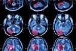
Functional neuroimaging techniques (single photon emission computed tomography [SPECT] and positron emission tomography [PET]) to investigate the nigrostriatal dopaminergic pathway at pre-synaptic and post-synaptic levels showed, in patients with two PINK1 mutations, a significant loss of pre-synaptic dopaminergic terminals in the putamen and caudate nucleus compared with normal controls, with normal post-synaptic function.6,8,14,18,23,43–46 Possible differences from idiopathic PD, not observed in all patients, include a more symmetrical reduction of dopamine re-uptake with less evident anteroposterior gradient and a slower progression of dopaminergic neuronal loss, suggesting a more uniform loss of nigrostriatal projections and the possible presence of compensatory mechanisms in PINK1-related parkinsonism.18,43 Functional neuroimaging studies in healthy heterozygous carriers also showed a mild but significant (20–30%) reduction of dopaminergic terminals, suggesting a role of heterozygous mutations in determining subclinical abnormalities in clinically asymptomatic subjects.12,18,43
Transcranial ultrasonography of the substantia nigra in two homozygous and one heterozygous patient showed a hyperechogenic area significantly larger than in healthy controls although smaller than in idiopathic PD cases, in line with results observed in other monogenic forms of parkinsonism.19 Moreover, a few studies assessed the myocardial (123)metaiodobenzylguanidine (MIBG) uptake (correlating with the integrity of sympathetic cardiac innervation, usually compromised in idiopathic PD) in five patients with homozygous PINK1 mutations, obtaining results ranging from normal (n=3) to markedly decreased values.21,45,47 MIBG uptake was reported to be as low as in idiopathic PD in three affected carriers of heterozygous PINK1 mutations.21
The integrity of the somatosensory system was investigated in PINK1 homozygous and heterozygous patients and in healthy heterozygous carriers using a psychophysical method: the temporal discrimination paradigm. Temporal processing of tactile and visuotactile sensory stimuli resulted in variable alterations in all PINK1 mutation carriers, including not only homozygous patients but also healthy heterozygotes.48
Possible Role of PINK1 Heterozygous Rare Variants
Several hypotheses have been proposed to explain the finding of a single heterozygous mutation in a high proportion of PD patients undergoing PINK1 mutation screening, especially in sporadic cases with later age at onset.
First, it must be considered that a second mutation in the PINK1 gene could have been ‘skipped’ by the currently adopted screening techniques, not sensitive enough to detect all types of mutations. For instance, this could be true for large genomic rearrangements, mutations in the promoter, or intronic mutations causing abnormal splicing. However, several studies that tested for genomic rearrangements in the PINK1 gene have yielded negative results, giving evidence that these types of PINK1 mutation are extremely rare and surely not able to account for the large proportion of heterozygous cases. This also applies for the other two mutational mechanisms that, although occasionally described in some genes, are overall rare.
Second, the second mutation could reside in a different gene, in a hypothetic model of digenic/oligogenic inheritance. This was suggested by occasional reports, such as a family with heterozygous mutations in both the PINK1 and DJ-1 genes,49 and a sporadic patient compound heterozygous for PINK1 and Parkin mutations.50 However, this hypothesis was excluded in several screenings that failed to identify concurrent mutations in other known ARP genes or LRRK2 in PINK1 heterozygotes.17,20,21,23
A third hypothesis is that PINK1 heterozygous mutations could act as modifier factors of the parkinsonian phenotype, influencing the age at onset or clinical presentation. This was suggested by the occasional report of a heterozygous PINK1 mutation in patients with bi-allelic Parkin mutations, who presented psychiatric symptoms and an earlier age at onset compared with patients carrying the same Parkin mutations but wild type for the PINK1 gene.50
A possible oligogenic mechanism has also been suggested in a PINK1 homozygous patient with very early onset of parkinsonism, in whom sequencing of mitochondrial DNA also revealed two missense mutations in the ND5 and ND6 genes, encoding two subunits of complex I.51 However, the evidence in favor of this hypothesis remains scarce, and further studies are needed to explore this possibility.
Finally, there is growing consensus on the hypothesis that single heterozygous variants in PINK1, as well as in other ARP genes, could act as risk factors increasing susceptibility to development of PD in a multifactorial model of disease pathogenesis. This is mostly supported by functional neuroimaging studies (discussed above) and by a recent study of voxel-based morphometry applied to high-resolution structural MRI, also showing subclinical abnormalities in the nigrostriatal function of healthy heterozygous carriers.52 This hypothesis would also justify the occurrence of heterozygous variants also in unaffected controls and in individuals with very mild parkinsonian signs.13,14
A meta-analysis was recently conducted on published screens that analyzed the whole PINK1 gene in both parkinsonian cases and healthy controls, to evaluate whether the risk of developing PD is increased in heterozygous carriers.17 PINK1 heterozygotes appeared to be more frequent in cases than controls (1.7 versus 1.0%), with an odds ratio (OR) of 1.62 (95% confidence interval [CI] 0.88–2.99) that did not reach statistical significance (p=0.121). Thus, the contribution of PINK1 heterozygous rare variants to PD genetic susceptibility seems to be minor. Heterozygotes would have a less than two-fold increased risk compared with wild-type individuals, and the vast majority of them are likely to remain lifelong unaffected. However, in the absence of long-term follow-up studies and functional assays on heterozygous rare variants (especially missense changes), the significance of these variants in PD patients and their associated risk of developing PD in unaffected carriers are still poorly understood and largely unpredictable. ■


