touchNEUROLOGY touchNEUROLOGY
Multiple Sclerosis
Read Time: 6 mins

Biological Markers of Prognostic Value in Multiple Sclerosis

Copy Link
Published Online: Jun 4th 2011 European Neurological Review, 2008;3(2):94-96 DOI: http://doi.org/10.17925/ENR.2008.03.02.94
Authors: Franziska Di Pauli, Markus Reindl, Thomas Berger
Quick Links:
Article
Article Information
Article:

Multiple sclerosis (MS), a chronic inflammatory disorder of the central nervous system (CNS), is the most common neurological disease among young adults, and carries the potential risk of permanent disability. The pathological hallmarks of the disease are multifocal white (and, most recently, also grey) matter lesions, which are characterised by variable extents of inflammation, demyelination, axonal loss, gliosis and atrophy.1 MS has variable clinical presentations and highly heterogeneous disease courses, ranging from rare acute fulminate forms to benign MS without substantial disability. Eighty-five per cent of patients initially present with a clinically isolated syndrome (CIS); most of these patients go on to develop relapsing–remitting (RR) MS, with acute relapses alternating with periods of clinical remission or stability.2 Ultimately, more than half of (untreated) RRMS patients convert to secondary chronic progressive (SP) MS, which is characterised by accumulating neurological disability with or without superimposed relapses.3 The clinical outcome of MS is largely unpredictable for individual patients. The great variability of this complex disease highlights the need for reliable biological markers with high sensitivity and specificity that are able to predict the future disease course and treatment response. Furthermore, stratification of MS patients with regard to their dominating pathological processes would allow individualised differential therapeutic concepts. In this review, we discuss the prognostic value of biological markers that are currently under debate, including magnetic resonance imaging (MRI), cerebrospinal fluid (CSF) parameters and antibodies.

Markers to Predict Disease Progression
Magnetic Resonance Imaging as a Prognostic Marker

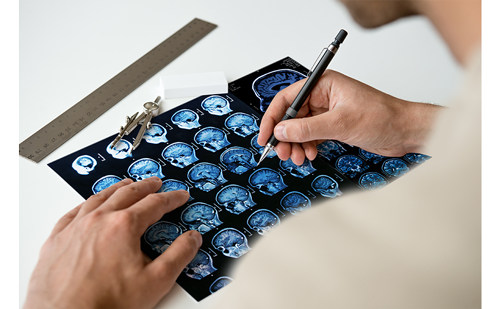
MRI is a well established tool for the diagnosis4 and management of MS that allows disease activity and progression to be monitored. The lesions detected on T2-weighted and gadolinium (Gd)-contrast-enhanced T1-weighted MRI reflect the pathological hallmark of the disease: the T2 lesion burden seems to be correlated with the number of preceding relapses5 and the use of Gd enables visualisation of blood–brain barrier disruption and therefore inflammatory disease activity.6 Thus, MRI has become a relevant surrogate outcome marker in MS clinical trials. Doubtless, MRI has its greatest relevance in patients with a CIS: evidence of dissemination of MS lesions in space and time and the extent of MRI activity are robust predictors of a first relapse.7,8 However, since commonly used MRI techniques show only a weak association with future disability,9 their prognostic value is limited and they are not useful for predicting clinical outcome in individual patients.10 Poor clinico-radiological correlations may be due to either insensitive clinical rating scales or methodological difficulties in the detection of pathological alterations, especially axonal damage, within the normal-appearing white (NAWM) and grey matter (NAGM).10,11 Neuropathology demonstrates that axonal loss, which seems to be the substrate of accumulating disability, occurs not only in classic MS plaques but also in NAWM and the cortex. Imaging of axonal loss and further brain atrophy is not sufficiently reflected by conventional MRI techniques. Although it has been suggested that the degree of disability depends mainly on the extent of brain atrophy,13 until now it has not been commonly agreed upon as a prognostic marker. Newly emerging and innovative MRI techniques, such as higher-resolution imaging, brain volumetry, magnetisation transfer imaging and magnetic resonance spectroscopy, and the combination of these different imaging parameters, will be more predictive for disease progression in MS patients in the future.

Oligoclonal Bands

The qualitative and quantitative measurement of elevated immunoglobulins (IgG) in the CSF of MS patients is the only laboratory biomarker included in MS diagnostic criteria. Isoelectric focusing (IEF) is the best qualitative method for detection of oligoclonal bands (OCBs),14 and has a sensitivity higher than 95% in MS15 and a specificity generally considered to be more than 86%.14 The value of the presence of OCBs to predict future disability remains controversial.16,17
IEF also allows the detection of oligoclonal IgM bands,18 which seem to be predictive for a more severe disease course with a shorter time period to the next relapse, an earlier disease conversion to SPMS and a higher grade of disability.19 These results need further evaluation in prospective multicentre studies concerning both the methodical procedure and the prognostic specificity and sensitivity before IgM OCBs can be used as markers in clinical practice.

Antimyelin Antibodies

Antibodies directed against myelin-oligodendrocyte-glycoprotein (MOG), which is exclusively localised on the surface of myelin sheaths and oligodendrocytes,20 and myelin basic protein (MBP), which constitutes 30% of total central myelin protein,21 have been suggested to predict future disease progression in patients with a CIS.22 The results of several subsequent studies were conflicting and ranged from highly significant22–24 to significant in sub-analyses25,26 to not significant at all.27–29 The different studies were all performed with the same type of assay for antimyelin antibodies, i.e. immunoblotting, thus the inconsistent results are likely due to varieties among the study cohorts. Whether antimyelin antibodies will be useful for clinical practice remains to be established.

Neuromyelitis Optica

Neuromyelitis optica (NMO) is an inflammatory demyelinating disorder that selectively affects the spinal cord and optic nerves.30 NMO was generally regarded as a subtype of MS with a high risk of severe disability and mortality. Recently, the presence of NMO-specific autoantibodies, NMO IgG, was proved,31 which supports the hypothesis that humoral immunity plays an important role in the pathogenesis of NMO.32 Aquaporin-4, a water channel located in astrocyte foot processes at the blood–brain barrier, has been identified as target antigen.33 NMO IgG is the first antibody of diagnostic value in a demyelinating CNS disease and distinguishes NMO patients from those with classic MS and other inflammatory MS variants. Furthermore, detection of NMO antibodies in patients with recurrent optic neuritis or with initial occurrence of longitudinally extensive transverse myelitis seems to predict subsequent relapses.34,35 In future, this may render early identification of NMO patients possible, thus allowing a rapid start with specific therapies such as plasmapheresis36 or B-cell-selective treatments.37

Markers to Predict Treatment Response
Interferon-β

Interferon-β (IFN-β) is one of the first-line disease-modifying therapies in MS and significantly reduces clinical and MRI disease activity. However, only half of patients respond well.38,39 Therefore, the identification of biomarkers to predict treatment responses and failures would be of great value to individualise patient management.
Biological treatments are well known to induce to some extent antidrug antibodies, which are responsible for the decrease/blockade of treatment effects and the occurrence of adverse events. A significant percentage of IFN-β-treated MS patients develop neutralising antibodies (NAb) to IFN-β.40 NAb-positive patients show higher relapse rates and more disease activity on MRI than NAb-negative patients, which confirms the clinical importance of NAb.41 NAb titres are variable and may change over time. They usually appear in the first year of treatment,42 and their occurrence depends on the immunogenicity and route of administration of the IFN-β product, as shown by lower frequencies for intramuscular administration, for example.43 Several guidelines on the use of anti-IFN-β antibody measurements (e.g. by a European Federation of Neurological Societies Task Force)44 recommend NAb testing after 12 and 24 months of IFN-β treatment. In NAb-positive patients, a further NAb test after three to six months is needed to confirm NAB persistency. IFN-β therapy should consequently be discontinued in patients with persistent high NAb titres.44 This strategy allows the risk of treatment failure to be minimised, because high NAb titres clearly precede their adverse clinical consequences and patients can thus be switched to alternative treatment options. More recently, other strategies, such as genetic or genomic approaches, have tried to identify factors that allow prediction of treatment responses,45 e.g. determination of the immunogenicity of IFN-β with regard to the future risk of NAb development.46

Natalizumab

Natalizumab is a humanised monoclonal antibody that binds to very late activation antigen 4 (VLA-4), an α4β1 integrin, and thereby prevents the migration of leukocytes through the blood–brain barrier. NAb to natalizumab occur early (usually within three months) during treatment and are persistent in 6% of patients. These NAb increase drug clearance and competitively block active drug binding to VLA-4.47 Thus, persistent NAb to natalizumab antagonise the otherwise very good treatment effects on relapse rate and disease activity.47 Furthermore, persistent NAb are associated with more hypersensitivity adverse reactions, which also mainly occur within the first three months of treatment. Again, once testing for NAb to natalizumab is routinely used it will constitute a risk minimisation tool regarding hypersensitivity reactions and future treatment failures for individual patients.
Another severe adverse advent during treatment with natalizumab regards the risk of progressive multifocal leukoencephalopathy (PML), wh-ch has been estimated as one case per 1,000 natalizumab-treated patients over 18 months.48 Despite extensive studies, no prognostic marker could be identified that allows determination of the risk of PML in advance.49

Conclusion

The heterogeneity of MS in terms of clinical presentation, genetic background and pathological and immunological features requires reliable (differential) diagnostic and prognostic markers for individual counselling and therapeutic management. Numerous studies have tried to identify such a (panel of) biomarker(s) with high specificity and sensitivity to define patients according to their suggested immunological phenotype, to determine the prognosis of disease progression and to predict treatment responses. Some substantial progress can be noted, such as new MRI techniques, NMO-IgG antibodies or NAb to IFN-β or natalizumab. However, much more effort is necessary to reach the goal of prognostically valuable biological markers to anticipate future disease course and treatment response in individual patients. Emerging biotechnical methods and increasing insight into the underlying pathomechanisms will discover new biomarker candidates, which should, after careful validation, improve the perspective and management of MS patients. ■

References

  1. Noseworthy JH, Lucchinetti C, Rodriguez M, Weinshenker Bg, Multiple sclerosis, N Engl J Med, 2000;343:938–52.
  2. O'Riordan JI, Thompson AJ, Kingsley DP, et al., The prognostic value of brain MRI in clinically isolated syndromes of the CNS. A 10-year follow-up, Brain, 1998;121(Pt 3):495–503.
  3. Weinshenker BG, Bass B, Rice GP, et al., The natural history of multiple sclerosis: a geographically based study. I. Clinical course and disability, Brain, 1989;112(Pt 1):133–46.
  4. Polman CH, Reingold SC, Edan G, et al., Diagnostic criteria for multiple sclerosis: 2005 revisions to the “McDonald Criteria”, Ann Neurol, 2005;58:840–46.
  5. Molyneux PD, Filippi M, Barkhof F, et al., Correlations between monthly enhanced MRI lesion rate and changes in T2 lesion volume in multiple sclerosis, Ann Neurol, 1998;43:332–9.
  6. Bruck W, Bitsch A, Kolenda H, et al., Inflammatory central nervous system demyelination: correlation of magnetic resonance imaging findings with lesion pathology, Ann Neurol, 1997;42:783–93.
  7. Rocca MA, Agosta F, Sormani MP, et al., A three-year, multiparametric MRI study in patients at presentation with CIS, J Neurol, 2008;255:683–91.
  8. Tintore M, Rovira A, Rio J, et al., Baseline MRI predicts future attacks and disability in clinically isolated syndromes, Neurology, 2006;67:968–72.
  9. Kappos L, Moeri D, Radue EW, et al., Predictive value of gadolinium-enhanced magnetic resonance imaging for relapse rate and changes in disability or impairment in multiple sclerosis: a meta-analysis. Gadolinium MRI Meta-analysis Group, Lancet, 1999;353:964–9.
  10. McFarland HF, Barkhof F, Antel J, Miller DH, The role of MRI as a surrogate outcome measure in multiple sclerosis, Mult Scler, 2002;8:40–51.
  11. Barkhof F, The clinico-radiological paradox in multiple sclerosis revisited, Curr Opin Neurol, 2002;15:239–45.
  12. Trapp BD, Peterson J, Ransohoff RM, et al., Axonal transection in the lesions of multiple sclerosis, N Engl J Med, 1998;338: 278–85.
  13. Fisher E, Rudick RA, Cutter G, et al., Relationship between brain atrophy and disability: an 8-year follow-up study of multiple sclerosis patients, Mult Scler, 2000;6:373–7.
  14. Freedman MS, Thompson EJ, Deisenhammer F, et al., Recommended standard of cerebrospinal fluid analysis in the diagnosis of multiple sclerosis: a consensus statement, Arch Neurol, 2005;62:865–70.
  15. Kabat EA, Moore DH, Landow H, An Electrophoretic Study of the Protein Components in Cerebrospinal Fluid and Their Relationship to the Serum Proteins, J Clin Invest, 1942;21: 571–7.
  16. Koch M, Heersema D, Mostert J, et al., Cerebrospinal fluid oligoclonal bands and progression of disability in multiple sclerosis, Eur J Neurol, 2007;14:797–800.
  17. Amato MP, Ponziani G, A prospective study on the prognosis of multiple sclerosis, Neurol Sci, 2000;21:S831–8.
  18. Sharief MK, Thompson EJ, Intrathecal immunoglobulin M synthesis in multiple sclerosis. Relationship with clinical and cerebrospinal fluid parameters, Brain, 1991;114(Pt 1A):181–95.
  19. Villar LM, Masjuan J, Gonzalez-Porque P, et al., Intrathecal IgM synthesis in neurologic diseases: relationship with disability in MS, Neurology, 2002;58:824–6.
  20. Brunner C, Lassmann H, Waehneldt TV, et al., Differential ultrastructural localization of myelin basic protein, myelin/oligodendroglial glycoprotein, and 2’,3’-cyclic nucleotide 3’-phosphodiesterase in the CNS of adult rats, J Neurochem, 1989;52:296–304.
  21. Boggs JM, Myelin basic protein: a multifunctional protein, Cell Mol Life Sci, 2006;63:1945–61.
  22. Berger T, Rubner P, Schautzer F, et al., Antimyelin antibodies as a predictor of clinically definite multiple sclerosis after a first demyelinating event, N Engl J Med, 2003;349:139–45.
  23. Tomassini V, De Giglio L, Reindl M, et al., Anti-myelin antibodies predict the clinical outcome after a first episode suggestive of MS, Mult Scler, 2007;13:1086–94.
  24. Greeve I, Sellner J, Lauterburg T, et al., Anti-myelin antibodies in clinically isolated syndrome indicate the risk of multiple sclerosis in a Swiss cohort, Acta Neurol Scand, 2007;116: 207–10.
  25. Rauer S, Euler B, Reindl M, Berger T, Antimyelin antibodies and the risk of relapse in patients with a primary demyelinating event, J Neurol Neurosurg Psychiatry, 2006;77:739–42.
  26. Kuhle J, Lindberg Rl, Regeniter A, et al., Antimyelin antibodies in clinically isolated syndromes correlate with inflammation in MRI and CSF, J Neurol, 2007;254:160–68.
  27. Pelayo R, Tintore M, Montalban X, et al., Antimyelin antibodies with no progression to multiple sclerosis, N Engl J Med, 2007;356:426–8.
  28. Lim Et, Berger T, Reindl M, et al., Anti-myelin antibodies do not allow earlier diagnosis of multiple sclerosis, Mult Scler, 2005;11:492–4.
  29. Kuhle J, Pohl C, Mehling M, et al., Lack of association between antimyelin antibodies and progression to multiple sclerosis, N Engl J Med, 2007;356:371–8.
  30. Wingerchuk DM, Hogancamp WF, O’Brien PC, Weinshenker BG, The clinical course of neuromyelitis optica (Devic’s syndrome), Neurology, 1999;53:1107–14.
  31. Lennon VA, Wingerchuk DM, Kryzer TJ, et al., A serum autoantibody marker of neuromyelitis optica: distinction from multiple sclerosis, Lancet, 2004;364:2106–12.
  32. Lucchinetti CF, Mandler RN, McGavern D, et al., A role for humoral mechanisms in the pathogenesis of Devic’s neuromyelitis optica, Brain, 2002;125:1450–61.
  33. Lennon VA, Kryzer TJ, Pittock SJ, et al., IgG marker of opticspinal multiple sclerosis binds to the aquaporin-4 water channel, J Exp Med, 2005;202:473-477.
  34. Interferon beta-1b in the treatment of multiple sclerosis: final outcome of the randomized controlled trial. The IFNB Multiple Sclerosis Study Group and The University of British Columbia MS/MRI Analysis Group, Neurology, 1995;45:1277–85.
  35. Weinshenker BG, Wingerchuk DM, Vukusic S, et al., Neuromyelitis optica IgG predicts relapse after longitudinally extensive transverse myelitis, Ann Neurol, 2006;59:566–9.
  36. Keegan M, Pineda AA, McClelland RL, et al., Plasma exchange for severe attacks of CNS demyelination: predictors of response, Neurology, 2002;58:143–6.
  37. Cree BA, Lamb S, Morgan K, et al., An open label study of the effects of rituximab in neuromyelitis optica, Neurology, 2005;64:1270–72.
  38. Stone LA, Frank JA, Albert PS, et al., Characterization of MRI response to treatment with interferon beta-1b: contrastenhancing MRI lesion frequency as a primary outcome measure, Neurology, 1997;49:862–9.
  39. Waubant E, Vukusic S, Gignoux L, et al., Clinical characteristics of responders to interferon therapy for relapsing MS, Neurology, 2003;61:184–9.
  40. Deisenhammer F, Reindl M, Harvey J, et al., Bioavailability of interferon beta 1b in MS patients with and without neutralizing antibodies, Neurology, 1999;52:1239–43.
  41. Neutralizing antibodies during treatment of multiple sclerosis with interferon beta-1b: experience during the first three years. The IFNB Multiple Sclerosis Study Group and the University of British Columbia MS/MRI Analysis Group, Neurology, 1996;47:889–94.
  42. Sorensen PS, Ross C, Clemmesen KM, et al., Clinical importance of neutralising antibodies against interferon beta in patients with relapsing-remitting multiple sclerosis, Lancet, 2003;362:1184–91.
  43. Ross C, Clemmesen KM, Svenson M, et al., Immunogenicity of interferon-beta in multiple sclerosis patients: influence of preparation, dosage, dose frequency, and route of administration. Danish Multiple Sclerosis Study Group, Ann Neurol, 2000;48:706–12.
  44. Sorensen Ps, Deisenhammer F, Duda P, et al., Guidelines on use of anti-IFN-beta antibody measurements in multiple sclerosis: report of an EFNS Task Force on IFN-beta antibodies in multiple sclerosis, Eur J Neurol, 2005;12:817–27.
  45. Baranzini SE, Mousavi P, Rio J, et al., Transcription-based prediction of response to IFNbeta using supervised computational methods, PLoS Biol, 2005;3:e2.
  46. Hoffmann S, Cepok S, Grummel V, et al., HLA-DRB1*0401 and HLA-DRB1*0408 are strongly associated with the development of antibodies against interferon-beta therapy in multiple sclerosis, Am J Hum Genet, 2008;83:219–27.
  47. Calabresi PA, Giovannoni G, Confavreux C, et al., The incidence and significance of anti-natalizumab antibodies: results from AFFIRM and SENTINEL, Neurology, 2007;69:1391–1403.
  48. Yousry TA, Major EO, Ryschkewitsch C, et al., Evaluation of patients treated with natalizumab for progressive multifocal leukoencephalopathy, N Engl J Med, 2006;354:924–33.
  49. Goodin DS, Cohen BA, O’Connor P, et al., Assessment: the use of natalizumab (Tysabri) for the treatment of multiple sclerosis (an evidence-based review): report of the Therapeutics and Technology Assessment Subcommittee of the American Academy of Neurology, Neurology, 2008;71:766–73.

Further Resources

Share this Article
Related Content In Multiple Sclerosis
  • Copied to clipboard!
    accredited arrow-down-editablearrow-downarrow_leftarrow-right-bluearrow-right-dark-bluearrow-right-greenarrow-right-greyarrow-right-orangearrow-right-whitearrow-right-bluearrow-up-orangeavatarcalendarchevron-down consultant-pathologist-nurseconsultant-pathologistcrosscrossdownloademailexclaimationfeedbackfiltergraph-arrowinterviewslinkmdt_iconmenumore_dots nurse-consultantpadlock patient-advocate-pathologistpatient-consultantpatientperson pharmacist-nurseplay_buttonplay-colour-tmcplay-colourAsset 1podcastprinter scenerysearch share single-doctor social_facebooksocial_googleplussocial_instagramsocial_linkedin_altsocial_linkedin_altsocial_pinterestlogo-twitter-glyph-32social_youtubeshape-star (1)tick-bluetick-orangetick-red tick-whiteticktimetranscriptup-arrowwebinar Sponsored Department Location NEW TMM Corporate Services Icons-07NEW TMM Corporate Services Icons-08NEW TMM Corporate Services Icons-09NEW TMM Corporate Services Icons-10NEW TMM Corporate Services Icons-11NEW TMM Corporate Services Icons-12Salary £ TMM-Corp-Site-Icons-01TMM-Corp-Site-Icons-02TMM-Corp-Site-Icons-03TMM-Corp-Site-Icons-04TMM-Corp-Site-Icons-05TMM-Corp-Site-Icons-06TMM-Corp-Site-Icons-07TMM-Corp-Site-Icons-08TMM-Corp-Site-Icons-09TMM-Corp-Site-Icons-10TMM-Corp-Site-Icons-11TMM-Corp-Site-Icons-12TMM-Corp-Site-Icons-13TMM-Corp-Site-Icons-14TMM-Corp-Site-Icons-15TMM-Corp-Site-Icons-16TMM-Corp-Site-Icons-17TMM-Corp-Site-Icons-18TMM-Corp-Site-Icons-19TMM-Corp-Site-Icons-20TMM-Corp-Site-Icons-21TMM-Corp-Site-Icons-22TMM-Corp-Site-Icons-23TMM-Corp-Site-Icons-24TMM-Corp-Site-Icons-25TMM-Corp-Site-Icons-26TMM-Corp-Site-Icons-27TMM-Corp-Site-Icons-28TMM-Corp-Site-Icons-29TMM-Corp-Site-Icons-30TMM-Corp-Site-Icons-31TMM-Corp-Site-Icons-32TMM-Corp-Site-Icons-33TMM-Corp-Site-Icons-34TMM-Corp-Site-Icons-35TMM-Corp-Site-Icons-36TMM-Corp-Site-Icons-37TMM-Corp-Site-Icons-38TMM-Corp-Site-Icons-39TMM-Corp-Site-Icons-40TMM-Corp-Site-Icons-41TMM-Corp-Site-Icons-42TMM-Corp-Site-Icons-43TMM-Corp-Site-Icons-44TMM-Corp-Site-Icons-45TMM-Corp-Site-Icons-46TMM-Corp-Site-Icons-47TMM-Corp-Site-Icons-48TMM-Corp-Site-Icons-49TMM-Corp-Site-Icons-50TMM-Corp-Site-Icons-51TMM-Corp-Site-Icons-52TMM-Corp-Site-Icons-53TMM-Corp-Site-Icons-54TMM-Corp-Site-Icons-55TMM-Corp-Site-Icons-56TMM-Corp-Site-Icons-57TMM-Corp-Site-Icons-58TMM-Corp-Site-Icons-59TMM-Corp-Site-Icons-60TMM-Corp-Site-Icons-61TMM-Corp-Site-Icons-62TMM-Corp-Site-Icons-63TMM-Corp-Site-Icons-64TMM-Corp-Site-Icons-65TMM-Corp-Site-Icons-66TMM-Corp-Site-Icons-67TMM-Corp-Site-Icons-68TMM-Corp-Site-Icons-69TMM-Corp-Site-Icons-70TMM-Corp-Site-Icons-71TMM-Corp-Site-Icons-72