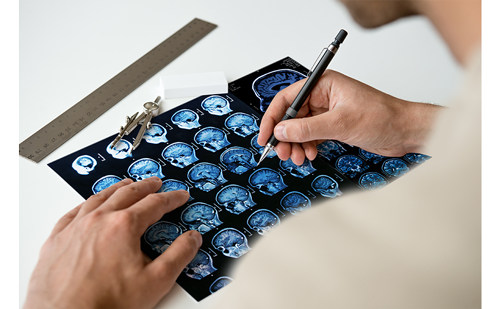
Recent advances in functional neuroimaging techniques have revolutionised the approach to neurosurgical planning. Functional magnetic resonance imaging (fMRI) techniques include blood-oxygen-level- dependent (BOLD) and diffusion tensor imaging (DTI) methodologies for non-invasively imaging brain activation and white matter fibres, respectively. In the last decade these techniques have evolved from purely research imaging tools used in cognitive neuroscience studies to clinically viable tools recognised by the medical community and regulatory bodies. These techniques hold considerable potential in the field of neurosurgical treatment planning.1,2
The goal of pre-operative treatment planning for tumour resection or seizure disorders is to differentiate eloquent cortex from structural brain lesions in close proximity, and to establish language lateralisation pre-operatively, in order to minimise surgical risk and post-operative neurological deficits. In addition to providing influential information regarding pre-operative risk assessment, the information provided by BOLD and DTI techniques is also critical to the neurosurgeon for developing intraoperative strategies and/or therapeutic approaches that otherwise might not be considered.
Procedures historically used as surgical mapping tools include the Wada test and direct electrical stimulation.3–6 However, these techniques are invasive and can impose some risk to the patients.
In the last few years, several prospective studies have been conducted with the goal of quantifying relative costs and benefits of using fMRI as an alternative pre-operative planning procedure, and evaluating concordance with currently used assessment tools. Studies directly comparing fMRI with Wada testing have demonstrated that fMRI imaging results significantly influence diagnostic and therapeutic decision-making, increase the confidence with which critical brain regions are identified and alter the surgical approach.7,8
Likewise, studies evaluating the effect of pre-operative fMRI localisation of language and motor areas on therapeutic decision-making show that treatment plans for nearly half of patients differ after fMRI assessment, surgical times are shortened, the spatial extent of resection is increased and craniotomy size is decreased, resulting in better outcomes and reduced risk of post-operative deficits.9 In addition, a recently published paper by Bizzi et al.10 compared fMRI with the current gold standard, intraoperative electrocortical mapping, and concluded that fMRI is a sensitive and specific method for mapping language and motor function.
Understanding the Application
Regardless of the application, using fMRI for pre-surgical mapping requires the evaluation of alternative imaging protocols to collect sufficient information to determine an optimal surgical approach. Imaging protocols differ in terms of their target brain region and functional complexity. All imaging protocols should include overt, measurable variables of behavioural performance in order to make unambiguous claims about associated neural activity.
During the early years of BOLD imaging, both in research and in the development of clinical applications, software for stimulus delivery and analysis of BOLD imaging data was developed by and for researchers and was often available as freeware. Solutions on the hardware side – for stimulus presentation (auditory or visual), response collection and synchronisation of stimulus delivery and image acquisition – were not provided by one vendor, but were often developed in-house. Multidisciplinary teams of researchers (biophysicist, neuropsychologist, research assistant, statistician, etc.) were required to integrate and implement solutions largely based on the availability of components from multiple vendors and the experience of the scientists. No US Food and Drug Administration (FDA)-approved, CE-marked, commercially available fMRI system existed that could be implemented solely by the MR technologist in the clinical setting. Fortunately, this situation has changed.
nordic fMRI Solution
NordicNeuroLab’s functional imaging solutions have been designed with the end user in mind, and the technology and solutions have continued to evolve with the needs of our customers, who span leading research institutions, medical centres, outpatient imaging centres and MRI system manufacturers. An important aspect of producing high-quality fMRI results involves standardisation of the activities performed by the MR technologist. NordicNeuroLab maintains a strong belief that the adoption of fMRI as a clinical diagnostic procedure critically hinges on the availability of easy-to-use tools that promise to yield reliable, repeatable results for the radiologist, treating clinician and neurosurgeon. The ability to streamline the fMRI examination procedure and seamlessly integrate the technology into radiology department workflow is a necessity. This need motivated NordicNeuroLab/NordicImagingLab to develop the nordic fMRI Solution, the only in-house-developed hardware/software solution for simplifying and standardising clinical fMRI exams.
nordicAktiva is the software that provides stimulus presentation and workflow guidance to facilitate the evaluation and comparability of results across exams. The intuitive, instructional interface directs the user through the process of selecting and presenting instructions and stimuli to the patient. The interface comes with a library of ready-to-use clinical paradigms. This capability is then interfaced with the MR scanner for immediate synchronisation with image acquisition.
Our fMRI hardware is the second component of the nordic fMRI Solution (see Figure 1). Nordic’s MR-compatible hardware provides audio-visual stimulus presentation, image synchronisation and response collection capabilities that simplify the complex operations associated with performing fMRI exams. This fully integrated hardware solution provides much flexibility as it is compatible with MR scanners from all major vendors.
nordicICE, and now our new clinical software nordicBrainEx, optimise overall workflow and minimise processing times while incorporating advanced features such as enhanced 2D/3D visualisation of BOLD and DTI tractography, advanced region of interest capabilities and time-course evaluation, easier export of images to picture archiving and communications systems (PACS) and neuronavigation systems and an automated clinical report capability for generating customised reports.
Case Report
Following is a case report of neurosurgical removal of a low-grade glioma using fMRI–DTI fused images for neuronavigation close to the Wernicke’s area.
Presentation
A 29-year-old man was admitted to the Department of Neurosurgery following a grand mal convulsion. MRI and MR spectroscopy (MRS) revealed an astrocytoma-like tumour within the left middle temporal gyrus. No neurological deficit was found in the otherwise healthy young male patient. As the tumour was close to the Wernicke’s area, an fMRI–DTI-guided frameless navigated microsurgical removal was planned.
Data Acquisition and Image Processing for BOLD and Diffusion Tensor Imaging Exams
MRI was performed on a SIEMENS 3T Trio scanner with a 12-channel phased-array head coil. Three image series were performed: an fMRI (text reading), DTI for tracking fibres within the speech areas and T1 MP-RAGE for structural imaging. After surgical removal of the lesion, fMRI and structural imaging were repeated. NordicNeuroLab’s nordic fMRI Solution was used to conduct the fMRI exam.
All fMRI image processing was performed using nordicICE software (BOLD module). A standard general linear model (GLM) approach was used for statistical analysis. Both BOLD and DTI images were co-registered to the structural images collected during the session. Fibre tracking was performed using the DTI module of nordicICE. The multiplanar reconstruction mode was used for fusing the co-registered fMRI activation information and the DTI fibre data to the structural images. Speech centres were selected as seeds for the fibre tracking. Fibre tractography and BOLD data were saved and exported to surgical navigation (see Figure 2).
Conclusion
During general anaesthesia, a generous left temporal craniotomy was made and the tumour extensions onto the cortical surface were delineated using the navigation system. Tumour resection was performed by microsurgical techniques. Histology revealed World Health Organization (WHO) grade II or III astrocytoma. The postoperative period was uneventful. A control MRI–fMRI confirmed a complete resection (see Figure 2, top image, right side). Functional results were comparable to those of the pre-operative findings, and no aphasia was observed after surgery.
In summary, the use of fMRI–DTI fused structural images for neuronavigation can help substantially in surgical planning and risk assessment when tumours are located close to eloquent brain regions. Thus, brain functions can be preserved and awake craniotomies may be avoided. ■


