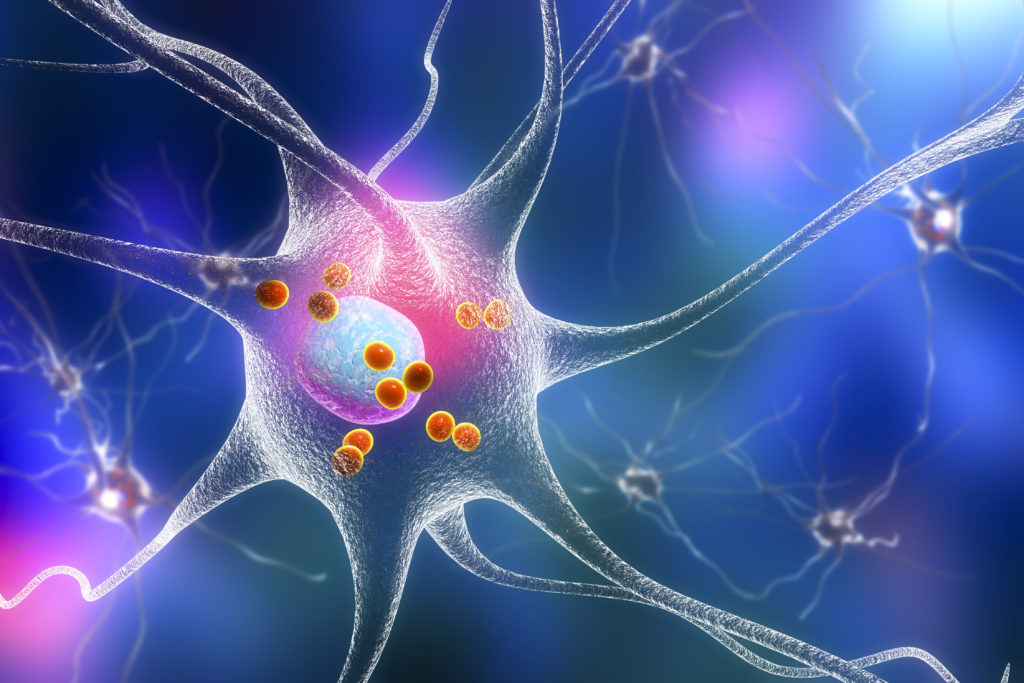
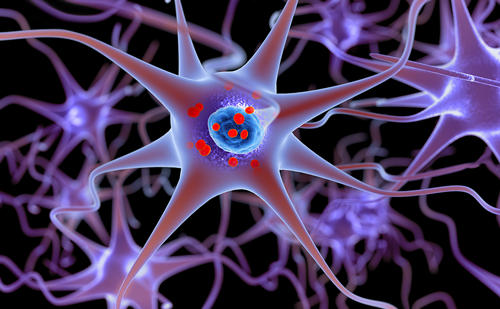
Parkinson’s disease (PD) is a progressive neurodegenerative disorder characterized by the loss of dopamine production in the substantia nigra. While oral therapies can effectively augment the dopaminergic pathway, their effect lessens as the disease progresses, and patients require multiple adjustments to their medication regimen. Neuromodulation using deep brain stimulation (DBS) is a powerful tool to ameliorate the symptoms of PD. A DBS system involves the implantation of electrodes into a neuroanatomical target. This delivers electrical impulses to specific brain areas to overcome abnormal neuronal activity in the stimulation target.1 The beneficial effects of DBS as compared with best medical therapy have been well described and it is currently a widely used treatment for most motor symptoms in PD.2–4 Dr Vanegas discusses her research on the use of imaging to predict outcomes of DBS.
Q. How are deep brain stimulation devices currently used in Parkinson’s disease?
PD is the second most common neurodegenerative disease after Alzheimer’s disease,5 and is characterized by both motor and non-motor symptoms. Non-motor symptoms include, but are not limited to, apathy, depression, anxiety, and fatigue, which are often more challenging to treat than motor symptoms. DBS is one of the most life-changing therapies for patients with PD, by specifically addressing their motor issues. However, we are less successful at treating non-motor, specifically neuropsychiatric symptoms, as these could worsen or even appear de novo after surgery. My research seeks to establish imaging predictors of motor and neuropsychiatric outcomes of DBS in patients with PD.
Q. Could you tell us a little about your current research?
My current research focuses on neuromodulation. The first line is trying to determine how imaging can help predict the outcomes of neuromodulation. We use high-resolution diffusion tensor imaging to evaluate our patients before they undergo DBS surgery in order to establish baseline brain characteristics. We then evaluate the connectivity of electrodes once patients have been treated with DBS.6 Our patient cohort is then followed over time using motor and non-motor measures. We hope to find pre-operative indicators of response that will predict the way in which patients will respond to the therapy. Our efforts have successfully found interesting observations on baseline structural connectivity that may end up being predictive of specific postoperative outcomes. The DBS leads have multiple contacts that we can use independently or in combination, at different frequencies and amplitudes of stimulation, therefore there is a lot of flexibility in the programming of these patients. While previous studies have focused on which contacts are most beneficial for motor symptoms,7 this study will focus on which contacts are most beneficial for cognitive symptoms over time. The next phase will be to investigate how the electrodes interact with the observed areas of degeneration. Ultimately, we hope to inform patient selection and DBS programming using brain imaging. This study is only in the preliminary stages; it is intended to last 5–10 years.
Q. How can imaging help you with the diagnosis of PD?
The diagnosis of PD is entirely clinical. DaTscan is a helpful imaging technique that provides information in regards to the presence of dopamine deficiency in the basal ganglia. DaTScan is a useful tool to distinguish Parkinsonian symptoms from different types of tremor or other neurodegenerative conditions, but it does not differentiate between different types of Parkinsonism. Although an abnormal DaTscan is not diagnostic of PD, it indicates abnormalities on dopaminergic uptake, which, in most cases, corresponds to PD.8 Research is still trying to identify a more reliable biomarker of PD. There are studies supporting the use of neuromelanin sequences on magnetic resonance imaging (MRI) for the diagnosis of PD but this has not yet been validated.9 There are also multiple studies using functional MRI and tractography that have correlated some aspects of the disease with imaging measures but those are not yet sufficient to make a diagnosis of PD.10
Q. Why is important to gather at-home data?
In order to improve the management of patients with PD, it will be necessary to understand the response to medication and how their daily activities are affected. For example, falls are a very common complication of PD and preventing those is important in order to control their morbidity and mortality. We require accurate information on the patient’s activities outside the office in order to optimize the treatment we provide. The use of questionnaires and diaries is challenging as this is subject to recall bias.11 In addition, some of our patients have cognitive issues and it may be difficult for them to complete these. It is important to get a better sense of how the disease is affecting our patients in order to develop new medications and neuroprotective approaches, in addition to optimizing current therapies.
Technology such as wearable sensors currently allows us to objectively measure patient’s movement and function during daily activities. These devices provide movement information as influenced by the patient’s natural environment and individual variability throughout the day.12
Q. Can you speak about the potential for DBS devices to also study PD?
Some DBS devices that we use to treat PD have the ability to record neuronal activity from the brain targets where they are implanted. Because symptoms of PD change throughout the day, especially during the effects of medications and during the presence of side effects such as dyskinesias, important research in DBS has focused on the potential use of closed loop stimulation.13 In other words, the information obtained from DBS devices in regards to neuronal activity during particular periods of the day, would allow us to customize the therapy that is being delivered to the brain. This will involve a large amount of data, as well as the integration with information obtained from imaging and other technologies, in order to create individualized programming algorithms. Thus, we hope that, in the future, DBS will be customized to the patient’s symptoms in real time. ⬛

