UCH-L1 Gene, Protein and Function
UCH-L1 Gene, Protein and Function
In 1983, a new human neuron-specific soluble protein was detected by high-resolution 2D electrophoresis, and was termed protein gene product 9.5 (PGP 9.5).1 The 27kDa protein, originally isolated from whole brain extracts, was used to raise specific antibodies that immunostained neurons and nerve fibres of the central and peripheral nervous system. It was observed that PGP 9.5 is highly abundant in the brain, representing up to 1–2% of all soluble brain proteins, which indicates an important role in the proper function of neurons.1 Subsequently, PGP 9.5 has been documented in segments of the renal tubules, spermatogonia, Leydig cells, ova, Merkel cells and dermal fibroblasts.2,3 In 1989 it was discovered that PGP 9.5 is a predominantly neuron-specific ubiquitin carboxyl-terminal hydrolase from the same family as the widespread and highly homologous UCH-L3.4,5 The gene contains nine exons which span over 10kb6 and is located on the short arm of chromosome 4.7 Enzymes from this thiol-protease family hydrolyse ubiquitin from the C-terminal end of substrates to generate the ubiquitin monomers, the active component of the cell’s ubiquitindependent proteolytic system, which degrades damaged proteins.4,5,8 A second function of UCH-L1 has since been described: as a dimer, UCH-L1 ubiquitinylates selective substrates in an ATPase-independent reaction.9 Additionally, it has been demonstrated that UCH-L1 regulates the degradation of free ubiquitin monomers in the cell.10
Parkinson’s Disease and UCH-L1
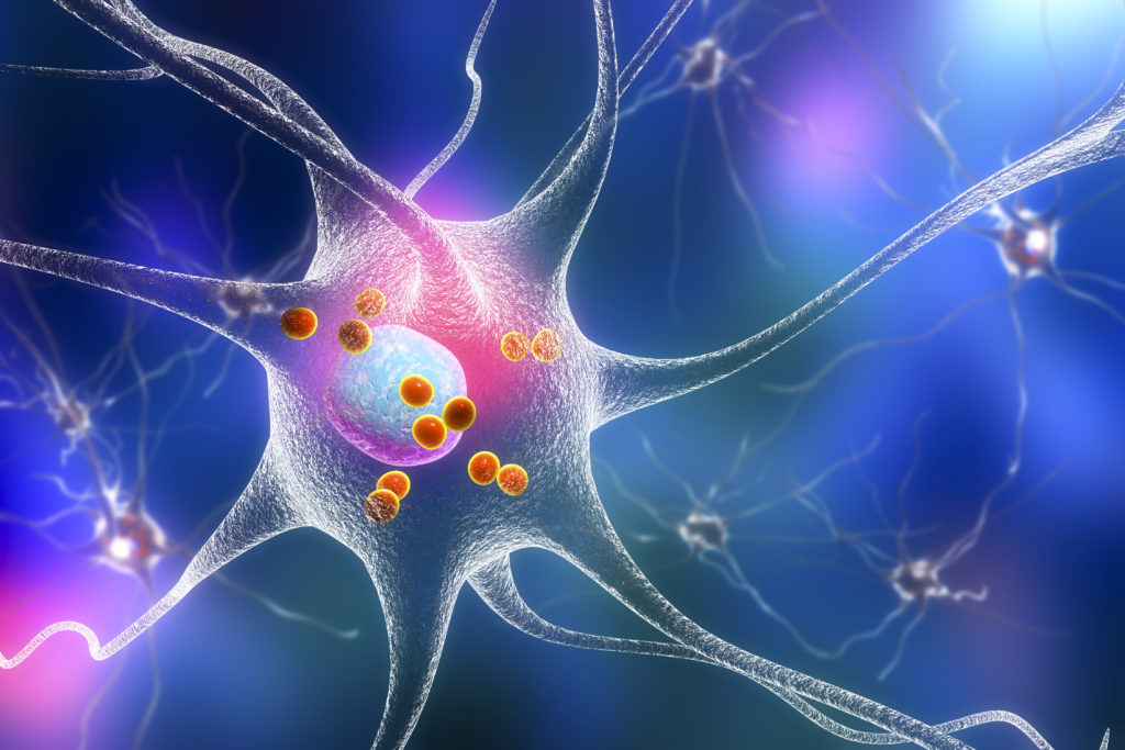
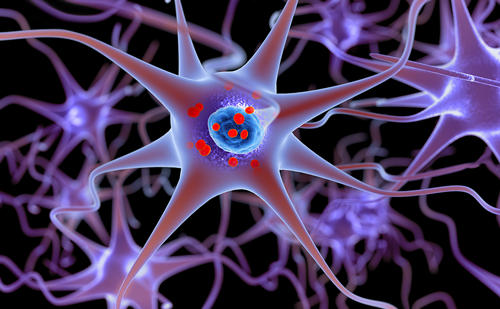
Parkinson’s disease (PD) is a major neurodegenerative disease that cannot be predicted and for which there is no cure. It affects more than 1% of the population over 65 years of age11 and is suggested to be somewhat more common among men than women. The incidence of PD is similar worldwide and the prevalence increases in proportion to regional increases in population longevity. The disease is characterised by bradykinesia, tremor at rest, rigidity and impaired balance. The main pathophysiological characteristics of PD are degeneration of dopamine neurons in substantia nigra and later in the ventral tegmental area in mesencephalon, but there is degeneration of other neurons as well. Another typical PD hallmark is the presence of Lewy bodies, which are proteinaceous intracellular inclusions mainly in the brainstem and cortex. UCH-L1 immunoreactivity has been detected in the cytoplasmic Lewy bodies within remaining dopamine cells in substantia nigra, making UCH-L1 a suitable candidate gene for PD.12 Downregulation of the full-length UCH-L1 protein form has been reported in post mortem brain from PD patients.13
UCH-L1 Mutations
In 1998, a candidate gene approach led to the discovery of the missense mutation I93M in exon 4 in a German family with autosomal dominant inherited PD.8 The coding region of the gene had been sequenced in 72 families with PD and the mutation in exon 4 of UCH-L1 was the only mutation discovered. Intensive research in different parts of the world failed to identify this or any other mutation in UCH-L1 in any other families or sporadic PD cases and raised the question of whether I93M is a truly pathogenic mutation.14 In vitro analyses from 1998 had indicated that the mutation causes a partial loss of hydrolytic function.8 A recent study analysing mutant transgenic mice expressing high levels of the human UCH-L1 I93M mutated enzyme under the PDGF-B promoter demonstrate a significant loss of dopamine neurons.15 However, the role of I93M in PD is inconclusive and needs further investigation.
In 1999, Lincoln et al. reported a polymorphism Ser18Tyr (S18Y) in exon 3 of UCH-L1.16 This genetic variant has been suggested to be protective since it was found to be inversely associated with sporadic PD.17 The protective effect of S18Y is dosage-dependent, indicating that a carrier of two Y alleles is less susceptible to developing PD than a carrier of one or no Y alleles. Several studies have addressed the role of this polymorphism in the past nine years, with variable results (for an overview, see Table 1). The inverse correlation with PD has been confirmed in nine studies, including a collaborative pooled analysis comprising 1,970 PD cases and 2,224 unrelated controls from 11 European sites.17-25
Eight further association studies failed to detect the inverse correlation of S18Y with PD, but there are no reports of the Y allele being a risk factor for PD (see Table 1).24,26–32 Interestingly, two of these studies claimed positive association when the material was stratified for early age at onset,26,31 although the reports were not corrected for multiple testing, which would nullify the significance of their finding, as pointed out by Healy et al. in 2006.27 Five of the studies reported a lack of association between S18Y and protection from PD regardless of stratification for age at onset.27–30,32 A large association study was initiated by Healy et al. in 2006 involving 1,536 PD patients and 1,487 controls of Caucasian origin from UK and Ireland.27 The S18Y variant was not protective against PD in any genetic model of inheritance (additive, recessive or dominant), and a haplotype-tagging approach did not detect other associated variants.27 The authors re-analysed the updated data from the earlier meta-analysis by Maraganore et al. from 2004 and drew the conclusion that there was no evidence that the S18Y variant of UCH-L1 exhibits protective effects in PD. Nevertheless, three more smaller association studies have been published on S18Y following Healy’s report that UCH-L1 is not a PD susceptibility gene, of which two are positive – one in a Chinese case-control study22 and one in a Swedish case-control study18 – and one is negative in a further Chinese PD study.32
There are a number of possible explanations for these conflicting results, the most important of which are the size of the case-control population analysed, the different geographical areas from which the material was collected and the different ways of analysing and stratifying the material for age at onset. The size of most case-control populations analysed ranges from 74 to 406 cases, except for the large Healy et al. study of more than 3,000 individuals (see Table 1). The earlier meta-analysis by Maraganore et al. showing a positive association has been criticised by Healy et al. for pooling data from small studies that all are influenced by small-study bias, particularly when stratifying for age at onset, and publication bias (small, negative studies remain more often unpublished).25,27 Importantly, Healy et al. show that exclusion from the Maraganore meta-analysis of a single Chinese study that does not follow the Hardy-Weinberg equilibrium results in loss of significance.21,25,27
The S18Y variant allele frequency differs considerable between geographical areas. The Y allele is relatively rare in the Caucasian population (~15–20%) and common in the Chinese (~50%) and Japanese (~40–50%) populations (see Table 1). All three studies on Japanese case-control populations report positive association for S18Y,20,21,24 whereas two of the studies on Chinese case-control populations report negative association and one a positive association. The two studies on case-control populations of Caucasian origin from the US reported conflicting results – one positive and one negative association – whereas in European case-control populations there seems to be a range from protective association in Northern and Central Europe (Sweden, Germany) to no association in other parts of Europe (France, Italy, the UK, Ireland). It is worth noting that only one of the S18Y studies was made on familial PD cases, and indicated no association.28
Healy et al. indicated that a small protective effect of the Y allele cannot be excluded,27 and the additional three association studies published – two positive and one negative – further show that genetic risk factors can vary between different populations.18,22,32 A locus found to be associated with or protective for disease in one population, such as Japan or Sweden, might also be in linkage disequilibrium with another, real protective locus that might not be linked in those ethnic populations where an association has not been found (for example the UK).
Does S18Y Influence the Enzymatic Activity of UCH-L1?
Position 18 of UCH-L1 is one of the few amino acid residues that is not conserved between humans and other mammals and has therefore been suggested not to be involved in the normal biological activity of UCH-L1,9 whereas the first identified mutation in PD, I93M in exon 4, causes a partial loss of hydrolytic function (~50%).8,33 In vitro studies with isolated proteins show that UCH-L1 exerts two opposing enzymatic activities that affect alpha-synuclein degradation: as a monomer, UCH-L1 can hydrolyse polyubiquitin chains, which promotes the ubiquitination and proteasomal degradation of, for example, alpha-synuclein; as a dimer, UCH-L1 ligates ubiquitin to certain proteins such as alpha-synuclein via an ATP-dependent K63 linkage, which spares it from proteasomal degradation. The I93M mutation inhibits the hydrolysation function and instead favours dimerisation, while the S18Y variant encodes a protein that is unable to dimerise and therefore favours degradation of proteins.9 Kyratzi et al. report that S18Y UCH-L1 comprises an antioxidant protective effect when expressed at physiological levels in human neuroblastoma cells or in primary cortical neurons, and that this property is not seen in UCH-L1-wild-type-expressing cells,34 further indicating a protective effect of S18Y. Transgenic mice overexpressing the S18Y protein variant have not been reported yet, whereas mice expressing human UCH-L1 with the I93M mutation show loss of dopamine neurons in substantia nigra15 together with a significant reduction of dopamine content in the striatum compared with non-transgenic animals. Interestingly, I93M transgenic mice also showed neuropathological changes such as silver-staining-positive argyrophilic grains in the perikarya of degenerating dopamine neurons and increased amounts of insoluble UCH-L1 in the midbrains.
Protective Variants in Other Diseases
Today only one other protective genetic variant has been described for PD – a haplotype of the AKT1 gene – but this finding has not been replicated so far.35 AKT1 is a serine/threonine protein kinase and its activation generates phosphorylation of several cellular proteins involved in the processes of apoptosis, metabolism and proliferation of neuronal cells.35 The mechanisms behind the protective haplotype are not yet known. There are several examples of protective variants reported in the literature for other diseases. One example is a mutation in the CCR-5 gene, called the Delta32 mutation, resulting in a modified protein unable to function as an HIV co-receptor and subsequently protecting homozygous carriers from being infected with HIV strains that require CCR-5 for penetrating cells.36 These results have been replicated by several groups. For disorders of the nervous system, few protective variants have been identified. Szolnoki et al. reported a cytoskeleton motor protein variant that may exert a protective effect on the occurrence of multiple sclerosis,37 but this study has not been reproduced so far. In Alzheimer’s disease the ε4 allele of the apolipoprotein E gene has been associated with an increased risk, as opposed to the minor ε2 allele, which has been suggested to have a protective effect against early-onset Alzheimer’s disease.38 In PD, the S18Y variant of UCH-L1 is the most studied example and the results are still controversial.
Conclusion
The abundance of UCH-L1 in the human brain, its presence in Lewy bodies and its involvement in protein degradation, a pathway disturbed in this neurodegenerative disorder, support the relevance of UCH-L1 involvement in PD pathogenesis. In vitro studies indicate that UCH-L1 exerts two opposing enzymatic activities that can affect the degradation of alpha-synuclein: the Y allele encodes a protein unable to dimerise, which favours degradation and reduces the risk of aggregation of synuclein in LB.9 This altered enzymatic activity strengthens the indication for the Y allele to be a protective factor for PD. The genetics of UCH-L1 is an example of how complex the evaluation of candidate genes can be for heterogeneous disorders such as PD. Association studies of the S18Y variant have led to inconsistent results in different populations, which may have several explanations. Even if the incidence of PD is geographically rather uniform, the importance of different genetic risk factors could vary between different populations. The locus found to be associated with decreased risk of PD in one material might be in linkage disequilibrium with another, real protective locus that is not linked in those ethnic populations where association has not been found.
The S18Y variant allele frequency differs strongly between geographical areas. The Y allele tends to be much more common in the Asian population compared with Caucasians, and a larger part of the studies on Asian case-control populations report positive associations with S18Y compared with the studies on Caucasian case-control populations. In European case-control populations there seems to be a trend from protective association in Northern and Central Europe (Sweden, Germany) to no association in other parts of Europe (France, Italy, the UK, Ireland). In the largest meta-analysis performed, Healy et al. found that neither the S18Y variant nor other genetic variants in UCH-L1 influence PD risk in Caucasians, but they admit that a small protective effect cannot definitively be excluded.27 So far there are no reports of the Y allele being a risk factor for PD. The protective effect of the Y allele seems to be more important for young age at onset since several groups report stronger association when stratifying for age at onset. Even groups reporting negative associations for the entire case-control population tend to find lower p-values when stratifying for age at onset.
In conclusion, further genetic and functional studies are needed to clarify the role of UCH-L1 in PD. A search for mutations in the promoter region that can affect messenger RNA (mRNA) or protein levels or in intronic regions affecting splicing is needed. It would also be interesting to develop transgenic mice expressing human UCH-L1 with the S18Y mutation and compare them with wild-type and transgenic mice with the I93M mutation, which show dopaminergic neuronal loss in substantia nigra.15 ■
Acknowledgements
We would like to acknowledge the Swedish Research Council, Hållstens Forskningsstiftelse, the Swedish Brain Foundation, the Swedish Parkinson Foundation, the Swedish Brain Power Initiative, USPHS grants and Karolinska Institutet Funds for the support of our research.

