Overview of Parkinson’s disease
Epidemiology
Parkinson’s disease (PD) is a common, chronic and progressive neurological condition. It is estimated to affect 100–180 people per 100,000 of the population and has an annual incidence of 4–20 per 100,000 people.1 The prevalence of PD rises sharply with age, from a mean of 41 per 100,000 in those aged 40–49 years to 1,903 per 100,000 in those aged over 80 years.2 The prevalence and incidence of PD is higher in men than in women.1
Risk factors for Parkinson’s disease
A number of putative risk factors for developing PD have been identified from systematic review and meta-analysis of observational studies. These include genetic and environmental risk factors, associated comorbidities and medication exposures, as well as early non-motor features that may represent the earliest stages of PD. The strongest risk factors associated with later PD diagnosis are having a family history of PD or tremor, and lack of smoking history. Other risk factors may include a history of mood disorder, exposure to pesticides, rural living, employment in farming or agriculture.3 Factors associated with the development of classic motor features of PD, which may represent an early pre-motor stage of the disease, include impaired sense of smell, sleep disturbances, rapid-eye movement (REM) behaviour disorder and constipation. A reduced risk of developing PD is associated with smoking, coffee-drinking, hypertension, and use of non-steroidal anti-inflammatory drugs (NSAIDs) or calcium (Ca2+) channel blockers.3

Clinical Features
The cardinal motor symptoms of PD are (see Figure 1):4
• bradykinesia (poverty/slowness of movement);
• rigidity;
• rest tremor; and
• postural and gait disturbance.
Patients with PD may present with all four cardinal symptoms or just one or two. However, a diagnosis of PD cannot be firmly made without features of bradykinesia with sequence effect, usually with asymmetry. Tremor is the presenting symptom in 70% of patients.5
Although PD is predominantly a disorder of motor function, patients with PD frequently develop non-motor symptoms. Non-motor symptoms are now recognised as a major determinant of quality of life and overall burden of disease.1 Non-motor symptoms in PD include:6
• sensory disorders (hyposmia and pain);
• autonomic dysfunction (orthostatic hypotension, neurogenic bladder disturbance, erectile dysfunction, constipation);
• neuropsychiatric symptoms (anhedonia, apathy, anxiety, depression, bradyphrenia, frontal executive dysfunction, dementia, psychosis); and
• sleep disorders (sleep fragmentation, reduced sleep efficiency, reduced slow-wave sleep, reduced REM sleep, REM sleep behavioural disorders, excessive daytime sleepiness, nocturnal akinesia/tremor, restless legs syndrome, periodic limb movements during sleep).
Non-motor symptoms were assessed in the Parkinson and Non Motor Symptoms (PRIAMO) study, a cross-sectional cohort of more than 1,000 patients with PD.7 In all, 98.6% of patients reported the presence of nonmotor symptoms, with a mean number 7.8 per patient (range 0–32). The most common were fatigue (58%), anxiety (56%), leg pain (38%), insomnia (37%), urgency and nocturia (35%), drooling of saliva and difficulties in maintaining concentration (31%). The frequency of non-motor symptoms increased in line with disease duration and severity (see Figure 2).


Diagnosis
The diagnosis of PD is primarily clinical, based on the history and examination.1 An example of a widely accepted diagnostic criteria is that developed by the United Kingdom Parkinson’s Disease Society (UK PDS) Brain Bank (see Table 1).8
In 2015, clinical diagnostic criteria for PD were published by the Movement Disorder Society (MDS).9 Again, the benchmark for these criteria is expert clinical diagnosis; the criteria aim to systematise the diagnostic process, to make it reproducible across centres and applicable by clinicians with less expertise in PD diagnosis. Although motor abnormalities remain central, increasing recognition has been given to non-motor manifestations; these are incorporated into both the current criteria and separate criteria for prodromal PD.9
The new MDS diagnostic criteria retain motor parkinsonism as the core feature of the disease, defined as bradykinesia plus rest tremor or rigidity, and include explicit instructions for defining these cardinal features. After documentation of parkinsonism, determination of PD as the cause of parkinsonism relies on three categories of diagnostic features: absolute exclusion criteria (which rule out PD), red flags (which must be counterbalanced by additional supportive criteria to allow diagnosis of PD), and supportive criteria (positive features that increase confidence of the PD diagnosis). The MDS criteria define two levels of diagnostic certainty: clinically established PD (maximising specificity at the expense of reduced sensitivity) and probable PD (which balances sensitivity and specificity).9
Disease progression and prognosis
PD is a chronic, progressive disorder for which there is presently no cure. Although PD typically presents with the cardinal motor symptoms, it is likely that the pathological process starts many years before these symptoms develop and it is likely they are reflected by the early appearance of non-motor features as described above.
Longitudinal assessment of newly diagnosed PD patients indicates that motor symptoms progress fastest in the early stages of disease.6 This is in agreement with pathological studies in PD patients that show an exponential decline in neurons in the substantia nigra over time.10
In a pathology study involving autopsy findings from 18 patients with PD or dementia with Lewy bodies, the average loss of neuronal density was 7% per year.10 A 29% loss was found to correlate with first motor symptoms and a 50% loss was present after five years of symptomatic disease. By extrapolation, the length of the presymptomatic phase was estimated at approximately five years.10
Recent studies have used non-biased stereologic counting methods to quantify substantia nigra pars compacta (SNc) neurons and striatal dopamine innervation.11 These demonstrate a complete loss of staining for dopamine terminals in the dorsal striatum within four years from initial diagnosis, suggesting completely degenerate or dysfunctional dopamine innervation by this time point.
The Sydney Multicenter Study of Parkinson’s Disease is one of the longest prospective studies, following 136 patients with PD from diagnosis (see Box 1).12,13 Long-term follow-up reveals the increasing burden of non-motor and non-dopaminergic symptoms as the disease progresses, with a very high prevalence of dementia by 20 years postdiagnosis. 12,13 Virtually all patients also experience levodopa-induced motor complications during this time period.
While disease progression in PD is inevitable, not all patients progress at the same rate. For example, patients with prominent tremor tend to progress at a much slower rate than those with the akinetic rigid form of the disease.14 Further, patients who have a mutation in the parkin gene generally have earlier onset and a slower course of PD.15 Currently, while the course of PD can be extended through treatment, progression cannot be halted.

Assessing the features of Parkinson’s disease
The multiple manifestations of PD can be assessed with the Unified Parkinson’s Disease Rating Scale (UPDRS), which was created in the 1980s and revised by the MDS-UPDRS Task Force in 2008.16 The MDSUPDRS consists of four parts (see Table 2) and the questions are designed to assess the clinical features of PD. All UPDRS questions have a response on a five-point scale (0=normal, 1=slight, 2=mild, 3=moderate, and 4=severe). The MDS-UPDRS total score (all parts) provides a comprehensive assessment of a patient with PD.
The examiner can also make a global assessment of the patient using the Hoehn and Yahr (H-Y) scale of clinical staging (see Table 3), which originated in the 1960s.17,18 The H-Y scale primarily captures physical disability and is widely used to establish eligibility for clinical studies and to track the progressive course of PD; its advantage is simplicity – patients are rated on a scale of 1 to 5. The disadvantage of the H-Y scale is that it does not capture comorbidities or the full spectrum of PD disability, such as depression, sleep disorders or dementia.18
Pathophysiology
An early pathologic hallmark of PD is a decline in dopamine-producing neurons in the SNc (see Figure 4).19 Neurologic signalling from this region of the mid-brain, through projection to the corpus striatum, is


integral to the regulation of normal movement. The striatum is the main input region of the basal ganglia for cortical information and plays an important role in motor control. Post-mortem pathology studies of PD patients’ brains show that even mildly affected patients have lost about 60% of their dopamine-producing neurons in the substantia nigra, particularly in the lateral portion.10 It is this loss, in addition to dysfunction of the remaining neurons, that accounts for the approximately 80% loss of dopamine in the corpus striatum in advanced patients.20
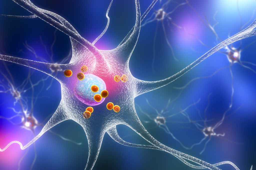
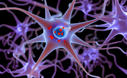
Another pathologic hallmark of PD is the presence of neuronal inclusions called Lewy bodies (see Figure 4).21 These structures contain misfolded aggregates of α-synuclein and ubiquitin. The Braak hypothesis suggests that α-synuclein inclusions may begin peripherally and spread to involve the mid-brain and then the cortex.22
Non-dopaminergic neurons are also involved in the pathology of PD and may account for many of the non-dopamine clinical features of the disease. Involvement can include neurons in the cerebral hemisphere, upper and lower brainstem, spinal cord, and peripheral autonomic nervous system, as well as those in the SNc. A raft of other non-dopaminergic neurotransmitter systems including acetylcholine, serotonin and norepinephrine neurons are also involved in the motor features of PD (see Figure 5).23
The precise mechanism accounting for the decrease in dopamineproducing neurons is not fully understood. It is generally thought that genetic and environmental factors may both play a role in causing neuronal death.24 While gene mutations account for as many as 15–20%


of cases, a genetic predisposition associated with an environmental trigger may be a far more common cause.6,24 The best documented and most widely investigated genetic causes are mutations in the LRRK2 (coding for leucine-rich repeat kinase 2), SNCA (coding for α-synuclein) and parkin genes.20


The current treatment paradigm in Parkinson’s disease
Symptomatic treatment for PD is typically initiated once the diagnosis is made or once motor symptoms become problematic. There is also much interest in drugs that might slow the rate of disease progression, and such a therapy, if it were established to be effective, would be started in all patients as soon as a diagnosis was made.
There are several possible approaches to the initial treatment of patients with PD (see Table 4).25 The choice is individualised, taking into account clinical and lifestyle characteristics and patient and physician preference. Monoamine oxidase-B (MAO-B) inhibitors are a reasonable first therapy because of their efficacy, good safety profile and potential disease-modifying effect.26 Others prefer to start with dopamine agonists because of their potential to avoid levodopa-induced dyskinesia; however, it is now clear that these agents can be associated with leg swelling, psychosis, sudden-onset sleep episodes, and impulse control disorders. Levodopa remains the most effective treatment for PD, but it is associated with the development of motor complications (motor


fluctuations and dyskinesia), although recent studies show that this risk can be markedly reduced with low doses.27 Regardless of what is chosen as initial therapy, most patients end up with polypharmacy, usually in low doses (see Figure 6 and Table 4).1,28,29 Other medication choices include amantadine and anticholinergics; however, these drugs are not particularly effective and are associated with cognitive impairment.25
With advanced disease, apomorphine infusion or penject, deep brain stimulation or intraduodenal levodopa infusion may be considered (see Table 5). However, it should be noted that these therapies are primarily designed to treat motor complications and do not provide anti-parkinsonian benefits that are superior to levodopa. Non-motor symptoms of the disease that may need to be managed include depression, sleep disorders, fatigue, psychosis/hallucinations, falls and dementia. Exercise/physiotherapy, disease education, occupational therapy, and speech and language therapy may all be helpful in individual patients.25
Unmet needs in mid- to late-stage Parkinson’s disease patients
The major unmet medical needs in the treatment of PD include:
• a dopaminergic therapy that provides the benefits of levodopa without worsening motor complications;
• a therapy for the non-dopaminergic features of PD such as falling and dementia; and
• a disease-modifying therapy that slows the rate of clinical progression.
Among these, motor complications appear to be the most readily treatable or preventable with available or future therapies. In early-

stage PD, dopaminergic treatment is extremely effective in controlling motor symptoms, with a single dose of levodopa providing benefit for many hours, despite the relatively short half-life of the drug. As the disease progresses, however, the beneficial motor response to levodopa gradually shortens (‘wearing off’) and dyskinesias increasingly complicate on-time (see Figure 7).30 Since the risk of developing both wearing-off and dyskinesias is increased by the use of high-dose levodopa, many physicians choose to use MAO-B inhibitors and/or dopamine agonists to delay the introduction of levodopa, and most eventually employ polypharmacy with low doses of levodopa combined with both of these agents.27
Motor complications – such as wearing-off and dyskinesias – are related to a variety of factors, including age, duration and dosage of levodopa, disease severity, gender and weight, as well as other pharmacokinetic and pharmacodynamic mechanisms.27,31 The majority of patients, particularly those with early disease, experience levodoparelated adverse effects after just a few years of treatment.32
The mechanism that is responsible for motor complications is not precisely known, but it is thought to relate to multiple factors including pulsatile delivery associated with administration of intermittent doses of standard levodopa.33 These in turn lead to altered gene expression in striatal input neurons, neurophysiologic alterations in pallidal output neurons and, ultimately, the development of motor complications. Current therapies for off periods include levodopa titration and supplemental doses of catechol-O-methyltransferase (COMT) inhibitors, dopamine agonists and MAO-B inhibitors. Attempts to control dyskinesia include titrating the dose of levodopa, and glutamatergic antagonists; amantadine is currently the only agent that provides antidyskinetic effects without worsening parkinsonism in double-blind studies, although it is associated with a number of adverse effects.34
Rationale for early add-on with safinamide to levodopa in mid- to late-stage Parkinson’s disease
Levodopa is the most widely used and most effective therapy for PD, but chronic treatment is associated with motor complications. The risk of developing motor complications is strongly linked to levodopa dose, independently of other predictive factors, including UPDRS measures of disease severity.27 Both dyskinesia and wearing-off become significantly more frequent at higher levodopa doses, particularly at doses greater than 400 mg/day (see Figure 8).27
In order to minimise the risk of motor complications in patients taking levodopa, it has been recommended that physicians should use the lowest dose of levodopa that provides satisfactory clinical control.27


This ‘levodopa-sparing’ approach may involve delaying the introduction of levodopa and/or maintaining lower doses of levodopa by adding-on therapies that are effective in improving PD symptoms to avoid the need to increase the levodopa dose. As a consequence, most PD patients ultimately receive a combination therapy with low-dose levodopa plus one or more other anti-parkinsonian agents.35 The goal is to provide good clinical efficacy with a reduced risk of side effects.35
A new approach to treating relatively early PD patients involves giving lowdose levodopa together with therapies that have multiple mechanisms of action, such as the recently European Medicines Agency (EMA)- approved drug safinamide (Xadago®).36 Safinamide is an oral therapy that is indicated for the treatment of adult patients with idiopathic PD as addon therapy to a stable dose of levodopa alone or in combination with other PD medicinal products in mid- to late-stage fluctuating patients.36 Safinamide has a dual mechanism of action that includes modulation of dopaminergic metabolism through selective, reversible inhibition of MAO-B, and blockade of voltage and use-dependent sodium (Na+) channels leading to inhibition of stimulated glutamate release.37
Safinamide
Chemical structure
Safinamide methansulfonate is the international proprietary name for the specific active enantiomer in safinamide. The International Union of Pure and Applied Chemistry (IUPAC) name for it is (S)-(+)-2-[4-(3-fluorobenzyl) oxybenzyl] aminopropanamide methanesulfonate. Safinamide is a member of the α-aminoamide chemical class. It is a small molecule that is chemically and metabolically stable and water soluble (see Figure 9).


Mechanism of action Safinamide acts through both dopaminergic and non-dopaminergic mechanisms (see Figure 10). Safinamide is a highly selective and reversible MAO-B inhibitor, leading to an increase in extracellular levels of dopamine in the striatum. Safinamide is also associated with state-dependent blockade of voltage-gated Na+ channels, resulting in modulation of calcium (Ca2+) channels and inhibition of stimulated release of glutamate.37,38
Monoamine oxidases (MAOs) A and B are mitochondrial bound isoenzymes that catalyse the oxidative deamination of monoamine neurotransmitters, such as serotonin, norepinephrine, dopamine and dietary amines. In the human brain, the MAO-B isoform is a key enzyme in the oxidative catabolism of dopamine; MAO-B inhibition blocks dopamine breakdown, increases synaptic dopamine levels, and enhances dopaminergic transmission in the brain. There is also evidence to suggest that MAO-B inhibition may have a diseasemodifying effect and may slow the rate of clinical progression.26,39.
Safinamide is a highly selective and potent MAO-B inhibitor. It is 1,000 times more selective for MAO-B than MAO-A in the human brain (see Figure 11), which precludes the need for any dietary restrictions.40 Nonselective MAO inhibitors and MAO-A inhibitors can lead to a potentially fatal hypertensive crisis known as the ‘cheese effect’, which results from failing to metabolise excess levels of dietary tyramine. Moreover, MAO-B inhibition with safinamide is completely reversible, thereby limiting potential drug–drug interactions and toxicity.40
Other dopaminergic mechanisms are relatively unaffected by safinamide. Safinamide does not display affinity for dopamine receptors and does not interfere with enzymes involved in the metabolism of levodopa: aromatic L-amino-acid decarboxylase (AADC) and COMT. Safinamide also has low affinity for 5-HT, glutamate, nicotinic, muscarinic and gamma-aminobutyric acid (GABA) receptors.41
Anti-glutamate activity
Safinamide can cause blockade of voltage- and use-dependent Na+ channels in activated neurons, leading to inhibition of excessive glutamate release.36 The Na+ channel inhibition is concentration- and state-dependent and does not influence physiological activity, thereby avoiding depressant effects on the central nervous system with the risk of side effects such as cognitive impairment and psychosis. Safinamide also does not affect L-type Ca2+ channels, which means that it has no effect on blood pressure or heart rate.40,42
Safinamide was originally studied as a putative anti-epileptic agent.43 In several central nervous system disorders, including epilepsy, schizophrenia and PD, neuronal over-excitation due to membrane depolarisation leads to Na+ channel opening. Profound depolarisation due to persistent Na+ channel opening leads to glutamate release and activation of voltage-dependent Ca2+ channels, allowing Ca2+ entry into the cell that triggers overactive release of glutamate.
When administered to rats, safinamide inhibited stimulated glutamate release in the hippocampus.44 This effect was dose-dependent and observed at levels proven to effectively block Na+ channels; importantly, however, safinamide did not affect basal glutamate release.44 Animal studies also suggest that safinamide has a differential impact on stimulated glutamate release in different regions of the basal ganglia complex. Specifically, when administered to rats, safinamide had no effect on stimulated glutamate release in the dorsolateral striatum, a partial inhibitory effect in the globus pallidus but near-total suppression in the substantia nigra pars reticulata and subthalamic nucleus.45
Safinamide preclinical development Pharmacokinetics and metabolism
Absorption
Safinamide is rapidly absorbed after single and multiple oral dosing, reaching Tmax within 1.8–2.8 hours post-dose under fasting conditions.36 Absolute bioavailability is high (95%), showing that safinamide is almost completely absorbed after oral administration and that first-pass metabolism is negligible.36 The high absorption classifies safinamide as a highly permeable substance.36
Distribution The volume of distribution (Vss) of safinamide is approximately 165 L, which is 2.5-fold of body volume, indicating extensive extravascular distribution.36 Total clearance was determined to be 4.6 L/hour, classifying safinamide as a low-clearance substance.36 Plasma protein binding of safinamide is 88–90%.36
Biotransformation
In humans, safinamide is almost exclusively eliminated via metabolism (urinary excretion of unchanged safinamide was <10%) mediated principally through high-capacity amidases, which have not yet been characterised.36 Safinamide’s metabolism is not dependent on cytochrome P450 (CYP)-based enzymes.36
Metabolite structure elucidation revealed three metabolic pathways of safinamide. The principal pathway involves hydrolytic oxidation of the amide moiety, leading to the primary metabolite ‘safinamide acid’ (NW-1153).36 Other metabolites include ‘O-debenzylated safinamide’ (NW-1199) and ‘N-dealkylated acid’ (NW-1689). None of these metabolites are pharmacologically active.36
Safinamide does not appear to significantly induce or inhibit enzymes at clinically relevant systemic concentrations.36 In vitro metabolism studies have indicated that there is no meaningful induction or inhibition of CYP450, CYP2A6, 2B6, 2C8, 2C9, 2C19, 2D6, 2E1 and 3A3/5 at concentrations which are relevant (Cmax of free safinamide 0.4 μM at 100 mg/day) in humans.36 Dedicated drug–drug interaction studies performed with ketoconazole, levodopa and CYP1A2 and CYP3A4 substrates (caffeine and midazolam) did not detect any clinically significant effects on the pharmacokinetics of safinamide, or levodopa, caffeine and midazolam.36
A mass balance study showed that the plasma area under curve (AUC) of the unchanged 14C-safinamide accounted for approximately 30% of the total radioactivity AUC0–24H, indicative of an extensive metabolism.36
Transporters
Preliminary in vitro studies have shown that safinamide is not a substrate for the transporters P-gp, BCRP, OAT1B1, OAT1B3, OATP1A2 or OAT2P1.36 Metabolite NW-1153 is not a substrate for OCT2, or OAT1, but it is substrate for OAT3. This interaction has the potential to reduce the clearance of NW-1153 and increase its exposure; however, the systemic exposure of NW-1153 is low (1/10 of parent safinamide), and as it is metabolised to secondary and tertiary metabolites, it is unlikely to be of any clinical relevance.36
Safinamide transiently inhibits Breast Cancer Resistance Protein (BCRP) in the small intestine.36 This could lead to clinically relevant drug–drug interactions with medicinal products that are substrates for BCRP and have a Tmax of ≤2 hours. At concentrations of 50 μM, safinamide inhibited organic anion-transporting polypeptide 1A2 (OATP1A2) and OATP2P1.36 The relevant plasma concentrations of safinamide are substantially lower, therefore, a clinically relevant interaction with co-administered substrates of these transporters is unlikely.36
Linearity/non-linearity
The pharmacokinetics of safinamide are linear after single and repeated doses. No time-dependency was observed.36
Elimination
Safinamide undergoes almost complete metabolic transformation (<10% of the administered dose was found unchanged in urine).36 Substance-related radioactivity was largely excreted in urine (76%) and, to a lesser extent, in faeces (1.5%) after 192 hours. The terminal elimination half-life of total radioactivity was approximately 80 hours.36 The elimination half-life of safinamide is 20–30 hours. Steady-state is reached within one week.36
Patients with hepatic impairment
Safinamide exposure in patients with mild hepatic disease increased marginally (30% in AUC), while in patients with moderate hepatic impairment exposure increased by approximately 80%.36 The drug should thus be used with some caution in patients with hepatic insufficiency.
Patients with renal impairment
Moderate or severe renal impairment did not alter the exposure to safinamide, compared to healthy subjects.36
Preclinical safety data
Retinal degeneration was observed in rodents after repeated safinamide dosing, resulting in systemic exposure below the anticipated systemic exposure in patients given the maximal therapeutic dose.36 No retinal degeneration was noted in monkeys despite higher systemic exposure than in rodents or in patients at the maximum human dose.36
Long-term studies in animals have shown convulsions (1.6 to 12.8 times human clinical exposure, based on plasma AUC).36 Liver hypertrophy and fatty changes were seen only in rodent livers at exposures similar to humans. Phospholipidosis was seen mainly in the lungs in rodents (at exposures similar to humans) and monkeys (at exposures greater than 12-fold higher than human).36
Safinamide did not present genotoxic potential in in vivo and in several in vitro systems using bacteria or mammalian cells.36
The results obtained from carcinogenicity studies in mice and rats showed no evidence of tumorigenic potential related to safinamide at systemic exposures up to 2.3 to 4.0 times, respectively, the anticipated systemic exposure in patients given the maximal therapeutic dose.36
Fertility studies in female rats showed a reduced number of implantations and corpora lutea at exposures in excess of three times the human exposure. Male rats showed minor abnormal morphology and reduced speed of sperm cells at exposures in excess of 1.4 times the anticipated human exposure.36 Male rat fertility was not affected. In embryo-foetal developmental studies in rats and rabbits malformations were induced at safinamide exposures two- and three-fold above human clinical exposure, respectively. The combination of safinamide with levodopa/carbidopa resulted in additive effects in the embryofoetal development studies, with a higher incidence of foetal skeletal abnormalities than seen with either treatment alone.36
In a pre- and postnatal developmental rat study, pup mortality, absence of milk in the stomach and neonatal hepatotoxicity were observed at dose levels similar to the anticipated clinical exposure.36 Toxic effects on the liver and accompanying symptoms such as yellow/orange skin and skull, in pups exposed to safinamide during lactation are mediated mainly via in utero exposure, whereas exposure via the mother’s milk had only a minor influence.36
Safinamide dosing and administration
Treatment with safinamide should be started at 50 mg/day. This daily dose may be increased to 100 mg/day on the basis of individual clinical need.36 No dose adjustment is required for elderly patients, patients with renal impairment or patients with mild hepatic impairment. The lower dose of 50 mg/day is recommended for patients with moderate hepatic impairment.36
Safinamide has a low risk of drug–drug interactions and can be used safely without any dietary tyramine restrictions.36 No dose adjustment is required with concomitant use of CYP1A2 and CYP3A4 substrates.36 Safinamide may be used with selective serotonin reuptake inhibitors

at the lowest effective dose. Concomitant use of safinamide and fluoxetine or fluvoxamine should be avoided or, if concomitant treatment is necessary, these drugs should be used at low doses.36 Safinamide must not be administered along with other MAO inhibitors (including moclobemide) or pethidine.36
Safinamide clinical development in mid- to latestage Parkinson’s disease
Overview of phase III studies
The clinical efficacy of safinamide 50 mg/day and 100 mg/day as add-on therapy to levodopa in mid- to late-stage PD patients experiencing motor fluctuations was evaluated in two 24-week, multicentre, double-blind, placebo-controlled trials: study 016 and the SETTLE study.46,47 The longterm efficacy and safety of safinamide 50−100 mg/day in this patient population were evaluated in study 018, an 18-month, double-blind, placebo-controlled extension to study 016.48
Study 016/018 – rationale and design
Study 016 was a 24-week double-blind, placebo-controlled, randomised, multicentre, multinational, phase III trial comparing oral once-daily safinamide 50 mg/day (n=223), safinamide 100 mg/day (n=224) versus placebo (n=222) for 24 weeks (see Figure 12).48 The primary efficacy endpoint of study 016 was the change from baseline to Week 24 in mean daily total on-time with no or non-troublesome dyskinesia, as recorded by patients or their caregivers in a home diary recorded at 30-minute intervals. Secondary efficacy variables include total daily off-time, UPDRS scores; Clinical Global Impression–Change (CGI-C) and –Severity (CGI-S) ratings; Patient Global Impression–Change (PGI-C) ratings; Parkinson’s Disease Questionnaire (PDQ-39) scores; EuroQoL Five-Dimension (EQ-5D) scores; change in levodopa daily dosage; Dyskinesia Rating Scale (DRS) scores; and Cogtest PD Battery scores.48
On completion of the 24-week treatment period, patients continued on their randomised study medication in an 18-month double-blind, placebo-controlled extension study (study 018), except for those patients experiencing dose-limiting side effects or clinically significant worsening.48 The objective of study 018 was to assess the long-term efficacy and safety of safinamide as add-on therapy to levodopa in patients with PD and motor fluctuations.48 The primary endpoint of study 018 was the change in dyskinesia over 24 months, as indicated by total DRS score during on-time. Patients continued to take the same dose of safinamide that they took at the end of study 016; patients who were unable to tolerate safinamide 100 mg/day had their dose decreased to 50 mg/day. Fifty of the 594 patients who completed study


016 did not enter study 018, 65.8% of study 016 patients completed the whole two-year treatment period, and 80.9% of study 018 patients completed the 18-month extension period (see Figure 13).46,48
Study 016/018 – main efficacy results
On-time with no/non-troublesome dyskinesia
Safinamide was significantly superior compared to placebo for the primary endpoint of on-time with no/non-troublesome dyskinesia (see Figure 14).47,48
Off-time
Safinamide significantly reduced off-time compared with placebo at 24 weeks (primary endpoint of the 016 study), and this effect was maintained for two years (secondary endpoint of the 018 study).


At baseline, mean daily off-time was 5.2 hours in each group. At two years, daily off-time was reduced by 0.62 and 0.75 hours with safinamide 50 and 100 mg/day, respectively, versus placebo.48
Safinamide was also associated with a significant improvement in off-time following the first morning dose of levodopa, assessed from patient-completed daily diaries.46 At six months, the mean difference versus placebo in off-time change after the first morning dose of levodopa versus baseline was –0.5 hours with safinamide 50 mg/day (p=0.0031) and –0.6 hours with safinamide 100 mg/day (p=0.0011).46
Dyskinesia
Safinamide failed to meet its primary endpoint, a reduction in dyskinesia, in the 018 study. However, subjects were initially recruited based on having off-time, and not all subjects had dyskinesia. Therefore, it was a high hurdle to demonstrate a reduction in dyskinesia. Nonetheless, for patients who had moderate–severe dyskinesia (approximately 25%), there was a significant improvement in DRS scores over the course of two years of treatment.
In the 018 study, for the entire population of patients, DRS scores were reduced from baseline by 31% and 27%, with safinamide 50 and 100 mg/ day, respectively, versus 3% with placebo, but this was not significant.48 In a post-hoc analysis, safinamide 100 mg treatment effect was statistically significant versus placebo in the subgroup of patients with moderate– severe dyskinesia (DRS score >4) at baseline (see Figure 15).48 This contrasts with most other studies, which aim to show that a reduction in off-time is not associated with an increase in troublesome dyskinesia. In the 018 study, not only was the reduction in off-time not associated with any increase in troublesome dyskinesia, but for the subgroup that had moderate-to-severe dyskinesia there was a significant reduction in dyskinesia score. This is consistent with safinamide’s dual mechanism of action, in which the MAO-B inhibition component improves parkinsonian features and reduces off-time, while the anti-glutamate component has the potential to lead to a reduction in dyskinesias. Interestingly, levodopa dose changes during the 24-month study did not differ among treatment


groups (see Table 6). A post hoc analysis in the subgroup of patients with no reduction of the levodopa dose showed a statistically significant effect of safinamide 100 mg, suggesting that the antidyskinetic effect of safinamide seen in the subpopulation was independent of the levodopa dose reduction (see Figure 16).49
The beneficial effect of safinamide on dyskinesia is unlikely to be related to a reduced dopaminergic stimulation, as demonstrated by statistically significant effects of safinamide during off-time, as discussed above. Rather, as noted above, glutamate has been suggested to play a role in the development of dyskinesia.40 Thus, the favourable effect on dyskinesia in the long term may be explained by inhibition of state- and use-dependent Na+ channels and stimulated glutamate release that are thought to contribute to the development of dyskinesia.49


Study 016/018 – safety and tolerability
The overall safety profile of safinamide is based on the clinical development programme performed in over 3,000 subjects, of whom over 500 were treated for more than two years.36
Overall tolerability In the 016/018 study, safinamide at doses of 50 or 100 mg/day was generally well tolerated with an adverse event (AE) rate similar to that of placebo (see Table 7).8


The most common treatment-emergent adverse event (TEAE) in patients receiving safinamide was dyskinesia.36 It should be noted that dyskinesia can increase with the introduction of any dopaminergic therapy; in this study, dyskinesia was mostly seen at the initiation of therapy and was much less pronounced with continuous treatment. Dyskinesia led to discontinuation in very few patients (approximately 1.5%) and did not require a dose reduction in any patient.36 The incidence of new-onset dyskinesia was 12.7% with safinamide 50 mg/ day, 13.2% with safinamide 100 mg/day and 15.4% with placebo.48
No clinically relevant differences in vital signs, laboratory tests, ophthalmological evaluations and electrocardiograms were observed among treatment groups.48
Discontinuations and serious adverse events
In the 016/018 study, rates of serious TEAEs and discontinuations due to TEAEs were similar in the three treatment groups (see Table 8). There were no treatment-related deaths.48
The SETTLE study – rationale and design
The SETTLE study was a 24-week double-blind, placebo-controlled, parallel-group, randomised, multicentre, multinational, phase III trial comparing oral once-daily safinamide (n=274) with placebo (n=275) as add-on therapy to a stable dose of levodopa in


patients with PD and motor fluctuations (daily off-time ≥1.5 hours) (see Figures 16, 17 and 18). Safinamide was started at a dose of 50 mg and increased to 100 mg after two weeks. The study allowed the inclusion of patients taking other concomitant PD medications (except other MAO-B inhibitors) at a stable dose.47 The primary efficacy endpoint was the change from baseline to week 24 in daily on-time, defined as on-time without dyskinesia and/or non-troublesome dyskinesia, as assessed by the patient-completed daily diary cards (18 hours/day).47
SETTLE study – main efficacy results
Safinamide was superior compared with placebo for the primary endpoint of on-time with no or non-troublesome dyskinesia (see Figure 19).47 At 100 mg/day, safinamide significantly increased mean daily on-time without troublesome dyskinesia, by 0.96 hours compared with placebo. Among key secondary outcomes, safinamide significantly reduced offtime, by 1.03 hours compared with placebo (see Figure 20).47 Safinamide also significantly improved motor function (UPDRS III), quality of life (PDQ-39, EQ-5D) and showed global benefit (CGI). Early-morning offtime was also reduced.47
SETTLE study – safety and tolerability
Safinamide was generally well tolerated in the SETTLE study, as indicated by high study completion rates (~89%) and low rates of safinamide discontinuation due to TEAEs (~5%). Overall, 67.9% of the safinamide group and 69.1% of the placebo group reported at least one TEAE (see Table 9). Among the most frequently reported TEAEs (see Table 10), dyskinesia was more common in the safinamide group than in the placebo group (14.6% versus 5.5%). The incidence of TEAEs rated as severe was lower in the safinamide group than in the


placebo group (6.9% versus 9.1%). However, dyskinesia was reported as severe in five patients (1.8%) on safinamide, compared with one patient (0.4%) on placebo.47
Post hoc and Pooled Analyses
A number of post-hoc analyses have been performed on data from the 016, 018 and SETTLE studies, including analyses of pooled data. Some of the most interesting exploratory and hypothesis-generating findings from the post-hoc analyses – including data on motor symptoms, non-motor symptoms, activities of daily living (ADL) and quality of life – are summarised below.
UPDRS III
In the 016 study, both doses of safinamide were effective in improving motor symptoms. Between baseline and week 24, UPDRS III scores at 24 weeks were improved by 6.1 and 6.9 points with safinamide 50 mg and 100 mg, respectively, versus a reduction of 4.3 with placebo.46 UPDRS III scores remained significantly lower with safinamide versus placebo at two years (mean reduction from baseline versus placebo: –2.13, p≤0.001), indicating that safinamide is associated with a sustained improvement in motor control.48
Other cardinal symptoms of Parkinson’s disease
Post hoc analyses of the pooled data from the SETTLE and 016 studies indicate that safinamide 100 mg/day was significantly effective in improving the cardinal features of PD including bradykinesia, rigidity, tremor and gait, as evaluated by the motor items of the UPDRS III scores. This was observed despite patients being on stable doses of dopaminergic treatments (see Table 11).50

CGI-C
In the 016 and 016/018 studies, the proportion of patients who were rated by their physician as ‘much/very much improved’ on the CGI-C was significantly higher with safinamide 50 or 100 mg/day than with placebo (see Table 12).36
UPDRS II
Safinamide 100 mg/day resulted in a significant reduction in UPDRS II scores versus placebo at both 24 weeks and 24 months (see Table 13).36
PDQ-39
In a post hoc analysis of pooled data from the SETTLE and 016 studies, the PDQ-39 index score improved between baseline and week 24 significantly more with safinamide 100 mg/day than with placebo. The safinamide treatment effect was statistically significant versus placebo for five of the eight PDQ-39 domains (see Table 14).51
Safinamide also significantly improved quality of life when used as addon to levodopa over the long term.48 At 24 months, the mean difference versus placebo in total PDQ-39 score reduction versus baseline was –10.48 with safinamide 50 mg/day and –18.36 with safinamide 100 mg/day (p=0.0195) (PDQ-39 scores at baseline were 225 in the safinamide 50 mg/day arm, 229 in the safinamide 100 mg/day arm and 230 in the placebo group).48 Statistically significant improvements in PDQ-39 subscale scores were observed with safinamide 100 mg/day versus placebo in ADL, emotional wellbeing, communication and bodily discomfort.48
Pain
A post hoc analysis of pooled data from the SETTLE and 016 studies found that patients treated with safinamide 100 mg as compared with placebo were significantly less likely to require concomitant pain treatments at six months. Furthermore, safinamide-treated patients showed significantly greater improvements in five domains of the PDQ- 39 (namely: mobility; ADL; emotional wellbeing; communication; bodily discomfort) and on items of the PDQ-39 ‘bodily discomfort’ domain related to musculoskeletal and neuropathic pain.51
Mood
A post hoc analysis of data from the 016/018 studies suggest that safinamide 100 mg/day may be effective in improving wellbeing and mood, as assessed with the PDQ-39 ‘emotional wellbeing’ domain and the GRID Hamilton Rating Scale for Depression (GRIDHAMD). 52 Furthermore, over the two-year treatment period, significantly fewer patients receiving safinamide experienced depression as

an AE compared with patients receiving placebo.52 Since depression was an exclusion criteria for entry into the trials, the results suggest that mood deteriorates with the progression of PD and that safinamide has a beneficial impact on mood that is maintained over two years of treatment.52 It is hypothesised that the favourable effect of safinamide on mood may be explained by modulation of glutamatergic hyperactivity.52
Summary
PD is a common, age-related progressive neurological disease that is most prevalent in the elderly, but can be seen in patients of all ages. The cardinal features are slowness of movement (bradykinesia/ akinesia), rigidity, resting tremor and postural and gait disturbances.
Although predominantly a movement disorder, PD is also associated with a wide range of non-motor symptoms such as alterations in mood and cognition, which can be an important source of morbidity. The pathologic hallmark of PD is a decline in the number of dopamineproducing neurons in the substantia nigra but pathology can be widespread, involving the cortex, brain stem, spinal cord and peripheral autonomic nervous system. Other key features are proteinaceous inclusions called Lewy bodies.
Treatment of patients with early-stage PD focuses on amelioration of motor symptoms with dopaminergic therapies. Current clinical guidelines recommend an escalating approach, frequently using polypharmacy that includes MAO-B inhibitors, dopamine agonists and low-dose levodopa based on symptom severity and age.25 Low doses of levodopa are recommended to try to avoid motor complications, but most patients eventually develop these problems after chronic levodopa treatment.25
In patients with mid- to late-stage PD, add-on therapies to levodopa may be needed to control motor symptoms. Add-on therapies may also allow lower doses of levodopa to be used, potentially offering good clinical efficacy while delaying the onset of levodopa-induced motor complications (dyskinesias and fluctuations). Further important goals of therapy in mid- to late-stage disease are the relief of non-dopaminergic symptoms, non-motor symptoms and levodopa-induced motor complications. Achieving these goals can be extremely challenging.
Safinamide is an important new medicine in the therapeutic armamentarium for fluctuating patients with mid- to late-stage PD, despite optimised pharmacotherapy including levodopa.
Safinamide has a unique dual mechanism of action on the dopaminergic and non-dopaminergic systems. Its mechanisms of action include modulation of dopaminergic metabolism through selective, reversible inhibition of MAO-B, blockade of voltage and use-dependent Na+ channels, Ca2+ channel modulation and inhibition of stimulated glutamate release.
Safinamide has been evaluated in a comprehensive clinical study programme involving more than 3000 patients with mid- to late-stage PD, of whom more than 500 were treated for two years. In these studies, safinamide 50–100 mg/day demonstrated efficacy in the treatment of motor fluctuations and motor symptoms in stabilised levodopa patients (improving UPDRS III score, and motor complications, as indicated by a significant reduction versus placebo in the primary endpoint of ontime without troublesome dyskinesias). Safinamide treatment was also effective for improving other motor complications (off-time, early morning akinesia), motor symptoms, non-motor symptoms, ADL and quality of life. There was also a suggestion of reduction in dyskinesia in a post hoc analysis of a subpopulation of patients. These effects have been proved in the short term (six months) and maintained in the long term (24 months).
In the pivotal studies, safinamide 50–100 mg/day was generally well tolerated with an AE rate similar to that of placebo. Safinamide dosing is relatively simple, with a low risk of drug–drug interactions, little need for dose adjustment and no need for dietary tyramine restrictions. In conclusion, the clinical data suggest that safinamide could be an appropriate choice as a first-line add-on therapy to levodopa in patients with PD who are experiencing motor fluctuations.


